Research Article |
|
Corresponding author: Ursula Eberhardt ( ursula.eberhardt@smns-bw.de ) Academic editor: Maria-Alice Neves
© 2022 Ursula Eberhardt, Alejandro Kong, Adriana Montoya, Nicole Schütz, Peter Bartlett, Henry J. Beker.
This is an open access article distributed under the terms of the Creative Commons Attribution License (CC BY 4.0), which permits unrestricted use, distribution, and reproduction in any medium, provided the original author and source are credited.
Citation:
Eberhardt U, Kong A, Montoya A, Schütz N, Bartlett P, Beker HJ (2022) Not (only) poison pies – Hebeloma (Agaricales, Hymenogastraceae) in Mexico. MycoKeys 90: 163-202. https://doi.org/10.3897/mycokeys.90.85267
|
Abstract
The species of Hebeloma have been little studied in Mexico, but have received attention as edibles and in trials to enhance production of edible fungi and tree growth through inoculation of seedlings with ectomycorrhizal fungi. Here we describe three new species of Hebeloma that are currently known only from Mexico. These species belong to separate sections of the genus: H. ambustiterranum is a member of H. sect. Hebeloma, H. cohaerens belongs to H. sect. Theobromina, while H. magnicystidiatum belongs to H. sect. Denudata. All three species were collected from subtropical pine-oak woodland; all records of H. cohaerens came from altitudes above 2500 m. Hebeloma ambustiterranum is commonly sold in the local markets of Tlaxcala as a prized edible mushroom. An additional nine species are reported from Mexico, of which eight are new records for the country: H. aanenii, H. eburneum, H. excedens, H. ingratum, H. neurophyllum, H. sordidulum, H. subaustrale and H. velutipes. First modern descriptions of H. neurophyllum and H. subaustrale, originally described from the USA, are given here.
Keywords
barcodes, Basidiomycota, ectomycorrhizal fungi, edible fungi, 3 new species, type studies
Introduction
Arguably, the best recognized vernacular English name for the genus Hebeloma is poison pie, although this name is often reserved for H. crustuliniforme, and other species within the genus are qualified versions of this name, e.g. H. mesophaeum is the veiled poison pie and H. pusillum is the dwarf poison pie (https://www.britmycolsoc.org.uk/library/english-names, accessed 18 Nov 2021). The name poison pie suggests what is, certainly in Europe, believed to be true for all members of the genus: that they are poisonous, or even if they were not, all too easily mixed up with poisonous members of the genus. Collecting Hebeloma for human consumption is generally discouraged (
In Mexico, the main interest in Hebeloma from the local community was either in the context of edibility (e.g.,
We have not had the opportunity to examine the material used in the respective publications. Given the difficulty surrounding species concepts of this genus, the presence of these species in Mexico should be treated with caution. Both, with regard to the consumption of mushrooms and the inoculation of tree seedlings, it would be advantageous to have a clear understanding of the species involved and the morphological and molecular characters that define them to recognize or verify collections or strains.
To the best of our knowledge, Hebeloma are not included in commercial ectomycorrhizal fungi mixtures currently sold to enhance tree growth, but it is one of the few genera that have been used in numerous nursery trials and transplanting experiments (e.g.,
From the taxonomic side, the Hebeloma of North America have been largely neglected since the work of Hesler and Smith in the 1970s and 1980s (
Within this paper, we present a list of Hebeloma species we have found during analysis of herbarium collections from Universidad Autónoma de Tlaxcala (
Materials and methods
All the material studied were dried specimens from the Universidad Autónoma de Tlaxcala (

Collection sites of studied material. Scale bar 1000 km. The map was generated with QGIS version 3.16.15 using WGS84, EPDG: 4326 (QGIS Association, QGIS.org, 2022). Shapefiles were provided by the Database of Global Administrative Areas (GADM); Accessed April 2018 to March 2022.
Sequences were obtained from the dried basidiomes by direct sequencing. At least the ITS (barcode) locus was generated for all Mexican collections and, in a number of cases, additional loci were sequenced. Internal transcribed spacer sequences were generated following methods detailed in
Sequence alignments were done online in MAFFT using the E-INS-i option (
Alignments were made for sections including new or rediscovered species, i.e., for H. sect. Hebeloma, H. sect. Naviculospora, H. sect. Theobromina and H. sect. Velutipes, including loci that were known to facilitate species recognition in the respective section (
Distances between sequences were calculated from the alignments used for the ML analyses as p-distances with pairwise deletion of gaps in MegaX (
Details of morphological analyses were provided in
All microscopic analysis was carried out on dried material, using a Leica DMRZA2 microscope with a Leica DFC495 camera connected to a computer running Leica Application Suite (LAS) V4 software.
The basidiospores were first studied in Melzer’s reagent to assess the shape, degree of dextrinoidity, ornamentation and the degree of loosening of the perispore. For the assessment of the degrees of ornamentation (O0, O1, O2, O3, O4), of the loosening perispore (P0, P1, P2, P3) and for the dextrinoidity (D0, D1, D2, D3, D4), we used
The material was then examined in 5% KOH. Photographs were taken of the basidiospores and also of the cheilocystidia (and pleurocystidia if any were present) and basidia at ×500 and ×1000. Because of the complex shapes of the cheilocystidia four measurements were made: length, width at apex (A), width at narrowest point in central region (M), and maximum width in lower half (B). The measurements were given in this order, and an average value was calculated for each of these measurements. The average width of the cheilocystidium in the vicinity of the apex appears to be an important character in the separation of species within Hebeloma (
Each collection studied has a database record number associated with that collection (beginning HJB); we give these numbers as we intend to make the database publicly available. If no other herbarium abbreviation or herbarium accession number is given, the HJB number is also the collection number within H.J. Beker’s herbarium.
Species were identified considering morphological and molecular data. In cases in which molecular data were not conclusive (as e.g., for H. eburneum and H. velutipes, or could not be obtained, as for the type of H. subaustrale), species identification followed morphology. For species not discussed in detail here, please refer to species descriptions in
Results
It appears that all of the species found in our sample, other than Hebeloma mesophaeum, are new species records for Mexico. Fig.
The analysis of taxa from H. sect. Hebeloma (from Mexico H. ambustiterraneum, H. excedens and H. mesophaeum) included ITS, RPB2 and Tef1a data, and 67 collections from 13 species. Hebeloma sordescens (H. sect. Hebeloma) was used for rooting. Hebeloma ambustiterranum was monophyletic in all single locus results and received support in ITS (100/100%) and RPB2 (85/98%). Conflicts between ITS and the other two loci were observed in relation to the position of H. pubescens (p.p.) and H. subtortum (ITS with H. excedens, H. mesophaeum and H. psammophilum; RPB2 and TEF1a with H. colvinii and H. velatum [= H. dunense,

ML topology of concatenated ITS, RPB2 and TEF1a sequences of Hebeloma sect. Hebeloma. Branch support was obtained through 1000 replicates of SH-like approximate likelihood ratio tests and ultrafast bootstrap annotated SH-aLRT/ufb at the branches for ≥ 85% SH-aLRT and ≥ 95% for ufb support. Dotted lines indicate parts of the tree where conflicts between single locus results were observed. Hebeloma sordescens (H. sect. Hebeloma) was used for rooting. Collections indicated with * are types; clade names indicated by * include type sequences. Collections and species names in red refer to Mexican material.
The analysis for H. sect. Denudata (in Mexico H. aanenii, H. eburneum, H. ingratum, H. magnicystidiatum and H. sordidulum) was based on ITS, mitSSU V6 and V9 of 78 collections from 17 species. Hebeloma echinosporum and H. populinum (H. sect. Denudata, subsect. Echinospora) were used for rooting. In the ITS tree, H. magnicystidiatum was part of the H. sordidulum clade (90/–%), which was included in a weakly supported clade (90/–%) with all other members of H. subsect. Clepsydroida considered in the analysis. Neither of the mitSSU results contradicted this relationship with any support, but there were conflicts between the ITS and mitSSU results and between the two mitSSU results in relation to the limits of the subsections and the relationship of H. hiemale (H. subsect. Hiemalia) and H. subsect. Clepsydroida and H. subsect. Crustuliniformia. In spite of this, the alignments were concatenated. The resulting phylogenetic hypothesis (Fig.

ML topology of concatenated ITS, mitSSU V6 and V9 sequences of Hebeloma sect. Denudata. Branch support was obtained through 1000 replicates of SH-like approximate likelihood ratio tests and ultrafast bootstrap annotated SH-aLRT/ufb at the branches for ≥ 85% SH-aLRT and ≥ 95% for ufb support or by thick lines in the case that at least one of the support values is equal to or exceeds the limits. Dotted lines indicate parts of the tree where conflicts between single locus results were observed. Hebeloma echinosporum and H. populinum (H. subsect. Echinospora of H. sect. Denudata) were used for rooting. Collections indicated with * are types; clade names indicated by * include type sequences. Collections in red refer to Mexican material.
The Mexican collections of H. aanenii clustered with their conspecifics from other countries, while the Mexican collections of H. eburneum were not in the same clade as H. eburneum collections from other countries, both clades received some support, one by ufb, the other by SH-aLRT (see Fig.
The analysis for H. sect. Velutipes (in Mexico H. neurophyllum and H. velutipes) was based on ITS, RPB2, TEF1a and mitSSU V6 of 59 collections from 12 species. Hebeloma bulbiferum and H. sinapizans (H. sect. Sinapizantia) were used for rooting. Hebeloma neurophyllum received good support (95/95%) in the ITS result, and is paraphyletic in relation to H. erebium in the RPB2 and TEF1a results, and in relation to H. celatum in the mitSSU V6 result. In spite of a number of conflicts concerning interspecific relationships within H. sect. Velutipes—intraspecific conflicts were not detected—the different single locus alignments were concatenated. The alignment included 2670 positions. In the analysis of the concatenated dataset (Fig.

ML topology of concatenated ITS, RPB2 and TEF1a and mitSSU V6 sequences of Hebeloma sect. Velutipes. Branch support was obtained through 1000 replicates of SH-like approximate likelihood ratio tests and ultrafast bootstrap annotated SH-aLRT/ufb at the branches for ≥ 85% SH-aLRT and ≥ 95% for ufb support or by thick lines in the case that at least one of the support values is equal to or exceeds the limits. Dotted lines indicate parts of the tree where conflicts between single locus results were observed. Hebeloma bulbiferum and H. sinapizans (H. sect. Sinapizantia) were used for rooting. Collections indicated with * are types; clade names indicated by * include type sequences. Collections in red refer to Mexican material.
Hebeloma velutipes was paraphyletic in relation to the other member species of the H. velutipes complex clade (H. incarnatulum, H. leucosarx and H. subconcolor). The position of the Mexican collections of H. velutipes in a separate clade (97/99%) was only supported by the mitSSU V6 data.
The analysis for H. sect. Theobromina (in Mexico H. cohaerens) was based on ITS, MCM7 and RPB2 of 32 collections from nine species. Hebeloma sinapizans was used for rooting. Hebeloma cohaerens was supported by all three single locus analyses (96–97/95–100%) and received full (100/100%) support in the analysis of the concatenated data (2152 bp) (Fig.

ML topologies with branch support obtained through 1000 replicates of SH-like approximate likelihood ratio tests and ultrafast bootstrap annotated SH-aLRT/ufb at the branches for ≥ 85% SH-aLRT and ≥ 95% for ufb support. Collections indicated with * are types; clade names indicated by * include type sequences. Collections in red refer to Mexican material A concatenated ITS, MCM7 and RPB2 sequences of Hebeloma sect. Theobromina, rooted with H. sinapizans (H. sect. Sinapizantia) B ITS sequences of H. sect. Naviculospora, rooted with H. islandicum (H. sect. Naviculospora).
The analysis for H. sect. Naviculospora (in Mexico H. subaustrale) was based on the ITS of 24 collections of eight species and included 703 positions. Hebeloma islandicum, provisionally placed by
Taxonomy
For species described from Europe please refer to
Hebeloma ambustiterranum
Type
Mexico. Tlaxcala: La Malinche National Park, 19.2749°N, 97.9825°W, alt. approx. 2800 m, on burnt soil in coniferous woodland under Pinus montezumae and P. teocote, 8 Jul 2017, H.J. Beker HJB16802 (holotype
Diagnosis
The small ellipsoid, non-dextrinoid, almost smooth basidiospores (on average 8.0–10.2 × 5.6–6.5 µm) and at least 50 full length lamellae distinguish this species from all other known North American Hebeloma species and the ITS sequence differentiates this species from all other known species, worldwide.
Etymology
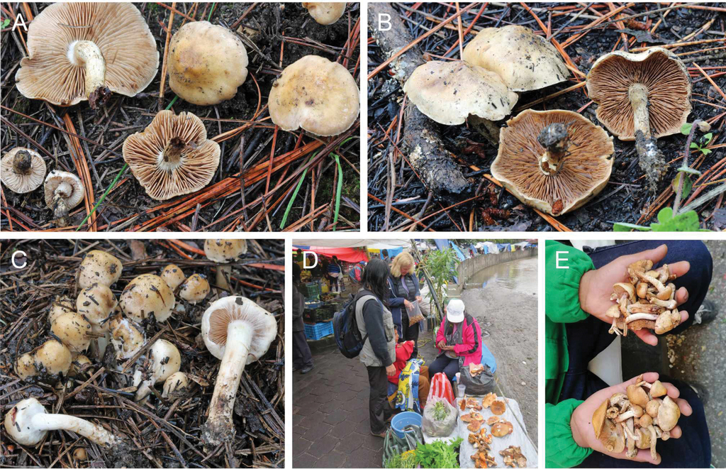
From ambustus (Latin adj.) meaning scorched, terra (Latin n.) meaning soil and the Latin suffix -anum indicating position to indicate growing on scorched soil. In Mexico, the local people burn the ground in the pine forests to encourage the growth of this mushroom, which they regard as an excellent edible mushroom. The local people refer to it in Nahuatl as the xolete de ocoxal (or ocoxalnanacatl), the mushroom of the pine needles from Chamusquinero, meaning from burnt ground.
Description
Pileus (12) 16–45 (52) mm diameter, usually umbonate or subumbonate, rarely convex or applanate; margin usually entire, sometimes involute particularly when young, often with remains of the universal veil, occasionally spotting, not hygrophanous; usually almost unicolored with color at center usually cream to ochraceous or clay-buff but may occasionally be darker, honey to sepia or umber, usually a little paler at the margin. Lamellae emarginate, white, cream to brown, with a weak white fimbriate edge sometimes visible and without droplets, number of full-length lamellae 50–74. Stipe (23) 24–60 (75) mm long, 3–8 (10) mm diameter at median, cylindrical, surface cream, ivory to pale brown but occasionally discoloring from the base upwards, sometimes strongly, fibrillose, at apex pruinose; base with white mycelium. Partial veil present on young specimens, whitish at first, before basidiospores mature, and often clear fibrils remaining on the stipe and pileus. Context in pileus white to cream, firm, in stipe stuffed, becoming hollow with age; taste not recorded, smell occasionally odorless but usually raphanoid, sometimes strongly so or with cacao components. Spore deposit color clay-buff.
Basidiospores based on n = 146 spores of the holotype, 5% to 95% percentile range 7.7–9.8 × 5.5–7.0 µm, with median 8.9 × 5.9 µm and av. 8.9 × 6.0 µm with S.D. length 0.68 µm and width 0.44 µm; Q value 5% to 95% percentile range 1.25–1.63, with median 1.48 and av. 1.47 with S.D. 0.11; spore size based on 33 collections medians 7.8–10.3 × 5.5–6.4 µm and av. 8.0–10.2 × 5.6–6.5 µm with av. S.D. length 0.61 µm and width 0.35 µm, av. Q 1.36 –1.61, ellipsoid or ovoid, with small apiculus, apex round or subacute, with a distinct thinning of the apical wall, guttulate with one or sometimes more oily drops, usually almost smooth even under immersion, with perispore not loosening, almost totally non-dextrinoid with just an indistinct brownish tint in Melzer’s reagent (O1; P0; D1); pale yellow to brown in KOH. Basidia 25–34 × 6–8 µm, with av. Q 3.7–4.4, cylindrical to clavate, hyaline, 4-spored. Cheilocystidium width near apex holotype 5% to 95% percentile range 3.5–5.3 µm, with median 4.3 µm and av. 4.3 µm with S.D. 0.63 µm; across 33 collections median 4.1–5.4 µm and av. 4.1–5.1 µm; examining approx. 20 selected cheilocystidia of each of the 33 collections yields a range for the avs. of 35–55 × 4.1–5.1 × 4.2–5.1 × 7.1–9.9 µm and 35 × 4.3 × 4.2 × 7.3 µm av. for holotype; av. ratios A/M: 0.96–1.15, A/B: 0.51–0.70, B/M: 1.55–2.31, mainly swollen in the lower half, some ventricose or lageniform, often with one or two septa, rarely geniculate or with some thickening of the median wall, hyaline. Pleurocystidia absent. Caulocystidia similar to cheilocystidia but more cylindrical and larger, up to 140 μm. Pileipellis an ixocutis; epicutis up to 100 µm thick, with gelatinized, often encrusted hyphae up to 6 µm wide; subcutis yellow and the trama below the cutis made up of cylindrical or occasionally ellipsoid cells up to 14 µm wide. Clamp connections present throughout the basidiome.
Ecology and distribution
In temperate coniferous woodlands on burnt ground with Pinus and Quercus. Growth habit usually scattered, rarely solitary or caespitose. To date, all collections of Hebeloma ambustiterranum recorded from Mexico at latitudes between 19°N and 20°N and altitudes above 2000 m.
Additional collections examined
Mexico. Mexico City: Municipality of Milpa Alta, approx. 19.1942°N, 99.0267°W, alt. approx. 2400 m, 4 Jul 2011, R. Vanegas-Enriquez (
Remarks
With its small ellipsoid, non-dextrinoid basidiospores and cheilocystidia swollen in the lower half, often lageniform or ventricose, this taxon clearly belongs to Hebeloma sect. Hebeloma and is closely related to the complex of species around H. mesophaeum. The close, but not crowded, lamellae with more than 50 full length lamellae rules out H. excedens and H. mesophaeum, both of which are widespread throughout North America (
Fig.

Holotype of Hebeloma ambustiterranum
Hebeloma cohaerens
Type
Mexico. Tlaxcala: Municipality of Panotla, 1 km al este de San Francisco Temezontla, approx. 19.3496°N, 98.2784°W, alt. approx. 2600 m, in deciduous woodland under Quercus sp., 23 Jul 2017, A. Montoya-Esquivel AME3102 (holotype
Diagnosis
The short clavate-ventricose cheilocystidia, with average apical width less 6.5 µm, the small (on average less than 10 × 5.5 µm), weakly ornamented but rather strongly dextrinoid basidiospores and the whitish to cream or buff color of the pileus, differentiate this species from other Hebeloma species.
Etymology
From cohaerens (adj. Latin) meaning united or joined together, to emphasize the connate habitus.
Description
Pileus (22) 32–38 (47) mm diameter, convex, often applanate, occasionally umbonate; margin smooth, often involute, particularly when young, occasionally eroded, not hygrophanous; usually almost unicolored, usually cream or buff, sometimes slightly paler towards margin. Lamellae often adnate or adnexed, occasionally emarginate, depth up to 4 mm, white, cream to brown, with white fimbriate edge but without droplets on the lamella edge, number of full-length lamellae 70–80. Stipe (31) 37–46 (48) mm long, (5) 7–8 (10) mm diameter at median, usually cylindrical but sometimes with a clavate base, surface cream, ivory, not discoloring, fibrillose, pruinose, particularly towards apex; base with white mycelium. Context in pileus and stipe white to cream, firm, in stipe stuffed; taste not recorded, smell earthy. Spore deposit color not recorded.
Basidiospores based on n = 64 spores of the holotype, 5% to 95% percentile range 8.6–10.5 × 4.9–5.7 µm, with median 9.4 × 5.3 µm and av. 9.5 × 5.3 µm with S.D. length 0.57 µm and width 0.26 µm; Q value 5% to 95% percentile range 1.64–1.95, with median 1.79 and av. 1.78 with S.D. 0.10; spore size based on four collections medians 9.1–9.5 × 5.3–5.6 µm and av. 9.1–9.5 × 5.3–5.5 µm with av. S.D. length 0.50 µm and width 0.30 µm, av. Q 1.65–1.78, amygdaloid, occasionally limoniform, with small apiculus and rounded apically, often subacute, with a distinct thinning of the apical wall and sometimes a papilla, usually guttulate with one or sometimes more oily drops, at most weakly ornamented (ornamentation only visible under immersion), with a perispore hardly loosening, rather strongly dextrinoid, becoming medium reddish brown in Melzer’s reagent (O1/2; P0; D3); yellow brown in KOH. Basidia 22–27 × 5–7 µm, with av. Q 3.7–3.8 µm, cylindrical to clavate, hyaline, 4–spored. Cheilocystidium width near apex holotype 5% to 95% percentile range 4.7–7.7 µm, with median 6.0 µm and av. 6.1 µm with S.D. 1.0 µm; across four collections median 5.6–6.4 µm and av. 5.5–6.3 µm; examining approx. 20 selected cheilocystidia of each of the four collections yields a range for the avs. of 33–36 × 5.5–6.3 × 3.5–4.1 × 5.5–6.6 µm and 33 × 6.1 × 4.1 × 6.5 µm av. for holotype. Cheilocystidium av. ratios A/M: 1.49–1.63, A/B: 0.86–1.03, B/M: 1.59–1.88, mainly clavate-ventricose, often with one or two septa. Pleurocystidia absent. Caulocystidia similar to cheilocystidia but larger, up to 90 μm long. Pileipellis an ixocutis with an epicutis up to 110 µm thick, with gelatinized, hyphae up to 6 µm wide; subcutis cream to pale yellow, and the trama below the cutis made up of cylindrical, often ellipsoid cells, up to 14 µm wide. Clamp connections present throughout the basidiome.
Ecology and distribution
In deciduous or mixed woodlands apparently associated with Quercus or Pseudotsuga. Growth habit mainly caespitose, sometimes with a few scattered basidiomes. To date, all collections of Hebeloma cohaerens recorded from Tlaxcala at altitudes of 2600 m or more.
Additional collections examined
Mexico. Tlaxcala: Municipality of Panotla, 1 km al este de San Francisco Temezontla, approx. 19.3496°N, 98.2784°W, alt. approx. 2600 m, in deciduous woodland under Quercus sp., 23 Jul 2017, A. Montoya-Esquivel (
Remarks
The small, short clavate-ventricose cheilocystidia, together with the small rather smooth, rather strongly dextrinoid basidiospores, support the placement of this species within Hebeloma sect. Theobromina. Within this section the pale cream to buff pileus color together with the caespitose habitus is unique.
The description was based on just four collections all from the same region of Mexico, and is not known from any other location. More records for this species will help to define better its morphological characters and its biogeographic preferences.
The minimum interspecific distance between the ITS sequences of H. cohaerens and sequences from other species is around 1.2%. The BLAST result of the type sequence of H. cohaerens against UNITE resulted in a hit of a soil sample sequence, pointing towards UNITE SH1563973.08FU (98.5% level). This SH includes two independently generated sequences from California (UDB0767851, soil sample,


Holotype of Hebeloma cohaerens
Hebeloma magnicystidiatum
Type
Mexico. Tlaxcala: Municipality of Totolac, Tepeticpac, 19.3457°N, 98.2226°W, alt. approx. 2400 m, on the ground in woodland under Pinus sp. and Quercus sp., 29 Aug. 1990, A. Estrada-Torres AET3093 (holotype
Diagnosis
The amygdaloid, non-dextrinoid, rather strongly ornamented spores with average Q value less than 1.6 and the capitate-stipitate cheilocystidia with average width at the apex greater than 9.5 µm distinguish this species from all other known Hebeloma species.
Etymology
From magni- (Latin, composite) meaning large and cystidiatus to emphasize the large capitate-stipitate cheilocystidia.
Description
Pileus 19–26 mm diameter, convex, surface dry, finely tomentose, cuticle separable, reddish yellow to brown in the center, and pale orange towards the margin. Lamellae emarginate, white, cream to orange brown as the spores mature, with a white fimbriate edge, and about 60 full-length lamellae. Stipe 10–21 mm long, 4–6 mm diameter at median, cylindrical, surface whitish but discoloring brown from the base upwards, with age or handling, fibrillose, at apex pruinose. Context in pileus white to cream, firm, in stipe stuffed, initially white to cream but becoming brown with age and handling, becoming hollow with age; taste fungal to sweet, smell raphanoid. Spore deposit not recorded.
Basidiospores based on n = 44 spores of the holotype, 5% to 95% percentile range 9.7–11.6 × 6.4–7.6 µm, with median 10.5 × 7.0 µm and av. 10.5 × 7.0 µm with S.D. length 0.62 µm and width 0.42 µm; Q value 5% to 95% percentile range 1.40–1.62, with median 1.49 and av. 1.50 with S.D. 0.07; amygdaloid, often limoniform, with small apiculus and rounded apically, often subacute to acute, with a distinct thinning of the apical wall and sometimes a clearly visible papilla, not guttulate, usually rather strongly ornamented, ornamentation visible even without immersion, with perispore at most somewhat loosening in a few spores, an indistinct brownish tint in Melzer’s reagent (O3; P1; D1); yellow-brown in KOH. Basidia 27.5–35 × 7.5–9 µm, with av. Q 3.9, cylindrical to clavate, without pigmentation, 4-spored. Cheilocystidium width near apex holotype 5% to 95% percentile range 6.1–14.3 µm, with median 9.1 µm and av. 9.7 µm with S.D. 2.61 µm; examining approx. 20 selected cheilocystidia of the holotype yields a range for the avs. of 55 × 9.7 × 7.3 × 4.3 µm av. and cheilocystidium av. ratios A/M: 2.58, A/B: 2.67, B/M: 0.95; mainly capitate-stipitate, unfortunately many collapsed in exsiccata. Pleurocystidia absent. Caulocystidia similar to cheilocystidia but larger, up to 80 μm long. Pileipellis an ixocutis; epicutis up to 110 µm thick, with gelatinized hyphae up to 7 µm wide; subcutis yellow; and the trama below the cutis made up of cylindrical or occasionally ellipsoid cells up to 17 µm wide. Clamp connections present throughout the basidiome.
Ecology and distribution
In woodland on the ground with Comarostaphylis and Quercus. Growth habit scattered. To date, with only one collection of this species, not possible to describe its distribution and ecology.
Remarks
With its amygdaloid, hardly dextrinoid basidiospores and capitate-stipitate cheilocystidia, morphologically this taxon clearly belongs to Hebeloma sect. Denudata and there to H. subsect. Crustuliniformia. The amygdaloid spores with rather small average Q value separate this species from all other studied Hebeloma from our database with more than 10,000 collections. While this may suggest that this is a rare species, we have insufficient Hebeloma collections from Mexico to reach such a conclusion. The single collection was collected in the 1990s, thus the only loci we could amplify were ITS and mitSSU variable regions V6 and V9.
The phylogenetic placement of H. magnicystidiatum within H. sect. Denudata is unresolved. As pointed out before (e.g. Eberhardt et al. 2016,

Holotype of Hebeloma magnicystidiatum
Hebeloma neurophyllum
Type
USA. New York: Coy Glen, Ithaca, approx. 42.4272°N, 76.5241°W, alt. approx. 125 m, on soil in woodland, 18 Oct 1906, N. Coil (holotype CUP-A-021514; isotype TENN-F-037531, HJB1000453, isotype WTU-F-039596, HJB1000558).
Diagnosis
Gregarium 7–8 cm altum, pileo 5–6 cm lato, stipite 5–6 mm crasso: Pileo ochraceo-cremeo vel fulvo-ochraceo, leviter viscido. Lamellis 8 mm latis, pallide cinnamomeo-rufis, late sinuatis, adnexis, costatis. Basidiis 4-sporis. Sporis subfusoideis, 12–15 × 7–8 µ[m]. Ad terram in silvis, Ithacae, N. Y. Stipite albo, fibroso-striato, cavo vel subfarcto.
English translation of diagnosis
Gregarious 7–8 cm high, pileus 5–6 cm broad, stipe 5–6 mm thick: pileus ochraceous-cream or fulvous-ochraceous, slightly viscid. Lamellae 8 mm broad, pale cinnamon-reddish, broadly sinuate, adnexed, intervenose. Basidia four-spored. Spores subfusoid, 12–15 × 7–8 μm. On the ground in woodland, New York. Stipe white, fibrous-striate, fistulose or almost stuffed.
Description
Pileus (26) 30–55 (60) mm diameter, convex, occasionally umbonate or broadly umbonate; margin often smooth, occasionally involute or wavy, not hygrophanous; usually unicolor, occasionally two colors, at center occasionally yellowish brown, ochraceous or cream, rarely fawn, cinnamon or clay-buff, sometimes slightly paler towards margin. Lamellae usually emarginate, occasionally adnexed, depth up to 9 mm, white, cream to brown, usually with white fimbriate edge, usually without droplets on the lamella edge but rarely some drops may be visible, number of full-length lamellae 70–94. Stipe (25) 31–75 (80) mm long, 5–14 (16) mm diameter at median, often clavate or bulbous, occasionally cylindrical, (7) 9–16 (18) mm wide at base, surface cream, ivory, rarely discoloring, occasionally velutinous, floccose or fibrillose, often pruinose, particularly towards apex. Veil not observed. Context in pileus white to cream, firm, in stipe usually hollow, rarely with superior hanging wick; taste mild, smell occasionally raphanoid or odorless, rarely fruity or earthy. Spore deposit yellowish brown to brownish olive.
Basidiospores based on n = 70 spores of the holotype, 5% to 95% percentile range 12.7–15.6 × 7.2–9.0 µm, with median 14.2 × 8.2 µm and av. 14.2 × 8.2 µm with S.D. length 0.93 µm and width 0.54 µm; Q value 5% to 95% percentile range 1.52–1.91, with median 1.74 and av. 1.73 with S.D. 0.12; spore size based on 47 collections medians 11.6–14.3 × 7.2–8.2 µm and av. 11.7–14.2 × 7.5–8.3 µm with av. S.D. length 0.898 µm and width 0.459 µm, av. Q 1.53–1.78, amygdaloid, usually limoniform, with small apiculus and rounded apically, often subacute to acute, with a distinct thinning of the apical wall and a clear papilla, occasionally guttulate with one or sometimes more oily drops, distinctly to strongly ornamented (ornamentation visible without immersion), with a perispore somewhat to distinctly loosening, at least in a few spores, strongly dextrinoid, becoming at least medium brown and often intensely red-brown in Melzer’s reagent (O3/4; P1/2; D3/4); yellow to brown in KOH. Basidia 20–43 × 7–10 µm, with av. Q 2.7–3.8 µm, cylindrical to clavate, with a median constriction, hyaline, 4-spored. Cheilocystidium width near apex holotype 5% to 95% percentile range 4.9–9.0 µm, with median 6.5 µm and av. 6.7 µm with S.D. 1.27 µm; across 47 collections median 4.5–6.8 µm and av. 4.6–6.7 µm; examining approx. 20 selected cheilocystidia of each of the 47 collections yields a range for the avs of 40–59 × 4.6–6.7 × 4.4–5.7 × 5.6–8.4 µm and 49 × 6.7 × 5.6 × 6.7 µm av. for the holotype. Cheilocystidium av. ratios A/M: 1.01–1.41, A/B: 0.68–1.23, B/M: 1.16–1.58, mainly gently clavate or ventricose, occasionally cylindrical, lageniform or clavate-lageniform or clavate-ventricose, often with one or two septa, sometimes clamped, often with plaques on the cystidial walls, occasionally geniculate or with basal wall thickening, rarely bifurcate, hyaline, rarely with yellow contents. Pleurocystidia absent. Caulocystidia similar to cheilocystidia but larger, up to 115 μm long. Pileipellis an ixocutis, epicutis up to 90 µm thick, with gelatinized, hyphae up to 6 µm wide; subcutis pale yellow to brownish yellow, and the trama below the cutis made up of cylindrical, often ellipsoid cells, up to 16 µm wide. Clamp connections present throughout the basidiome.
Habitat and distribution
Based on almost 50 collections, where only one possible associate was recorded, the most commonly recorded associates were Picea and Quercus, but Populus, Salix and Tilia were also recorded; the most commonly recorded families were Fagaceae, Pinaceae and Salicaceae, but Betulaceae and Malvaceae were also recorded. We have additional records where Alnus, Arctostaphylos, Betula, Dryas, Pinus and Polygonum were recorded as possible associates, but in each of these cases a number of possible associates were mentioned. All records of H. neurophyllum are from Northern America, where it is widespread across the region but primarily collected in temperate to boreal woodland, occasionally in urban areas.
Additional material examined
Canada. Alberta: Moose Hill, Breton, Edmonton, 53.1418°N, 114.6097°W, alt. approx. 810 m, on soil in mixed woodland under Picea mariana, 12 Aug 2017, H.J. Beker (HJB16856). Northwest Territories: Highway 3, between Yellowknife and Behchoko, 62.5198°N, 114.897°W, alt. approx. 165 m, on mossy soil in boreal, calcareous woodland roadside under Betula sp. and Salix sp., 7 Sep 2018, H.J. Beker, L. Davies (HJB18101). Yukon: Railway Station, Whitehorse, 60.7214°N, 135.0505°W, alt. approx. 665 m, on soil and litter in boreal shrubland riverside under Populus tremuloides and Salix sp., 31 Aug 2018, H.J. Beker, L. Davies (HJB17975). 3rd Avenue near Wood St intersection, Whitehorse, 60.7212°N, 135.0555°W, alt. approx. 665 m, on grassy, mossy soil in boreal urban roadside under Populus sp., 1 Sep 2018, H.J. Beker, L. Davies (HJB17981). MEXICO. Chihuahua: El Ranchito, approx. 28.3387°N, 105.4076°W, alt. approx. 1150 m, on soil in montane, subtropical woodland, 18 Aug 2001, A. Kong 3782 (
Remarks
With the mixture of gently clavate and ventricose cystidia alongside the strongly dextrinoid basidiospores, this species belongs within Hebeloma sect. Velutipes. Within this section the combination of spores with minimum average width 7.5 µm and a distinctly loosening perispore in at least some spores, together with the absence of pleurocystidia, defines this species. The collection of H. neurophyllum from Mexico, gathered at El Ranchito in Chihuahua, matches well with other collections of this species. We are not aware of any synonyms for this species.
In terms of ITS, the most similar to H. neurophyllum were H. celatum, H. erebium and H. quercetorum, the ITS sequences of which were around 99% similar (99.2–98.6%) to those of H. neurophyllum. Hebeloma neurophyllum appears to correspond to UNITE SH1733487.08FU (99%). Intriguingly, this species hypothesis includes a number of soil sample sequences from Estonia, suggesting that either H. neurophyllum occurs in Europe, too, or that species known to occur in Europe also contain ITS copies corresponding to H. neurophyllum below the detection limit of Sanger sequencing.


Hebeloma neurophyllum A basidiospores and B spore ornamentation of isotype TENN-F-037531 (HJB1000453) ×1600 C basidiospores of HJB17897 in Melzer’s reagent ×1600 D basidium of isotype ×1000 E–F cheilocystidia of isotype ×1000 G caulocystidia of HJB17975 ×500 H caulocystidia of HJB16856 ×500 I epicutis hyphae and J subcutis of isotype ×500. All in KOH, except C. Scale bars: 5 µm (A–F); 10 µm (G–J). Photos H.J. Beker.
Hebeloma subaustrale
= Hebeloma angustisporium Hesler, Kew Bulletin 32(3): 471 (1977)
= Hebeloma perangustisporium Hesler, Kew Bulletin 32(3): 478 (1977)
Type
USA. Florida: Gainesville, Alachua Co., approx. 29.651634°N, 82.324826°W, alt. approx. 50 m, on grassy, shady soil in lawn, 30 Oct 1941, G.F. Weber (holotype FLAS-F-19345, HJB1000402; isotype TENN-F-021177, HJB1000447).
Diagnosis
Pileo convexo-expanso, 3–4 cm. lato, subviscido, glabro, pallido-roseo, raphanico; lamellis sinuatis, latis, confertis; sporis subovoidcis, pallidis, levibus, 8–10 × 4–4.5 μ[m]; stipite acquali, pallido, 3 × 0.5 cm.
English translation of diagnosis
Pileus convex to applanate, 3–4 cm broad, slightly viscid, glabrous, pale pink, with raphanoid smell; lamellae sinuate, broad, crowded; spores subovoid, pale, smooth, 8–10 × 4–4.5 μ[m]; stipe equal, pale, 3 × 0.5 cm.
Description
Pileus (20) 32–45 (46) mm diameter, usually convex, occasionally umbonate; occasionally with remains of universal veil; margin often smooth, occasionally scalloped, not hygrophanous; usually unicolor, occasionally two colors, at center cream to buff to ochraceous, often becoming paler towards the margin. Lamellae usually emarginate, occasionally adnate or adnexed; white, cream to brown, usually with white fimbriate edge, without droplets on the lamella edge, number of full-length lamellae 80–92. Stipe 30–56 (70) mm long, 5–10 (11) mm diameter at median, often clavate or cylindrical, 5–13 (14) mm wide at base, surface cream, ivory to white rarely discoloring, pruinose, particularly towards apex. Context in pileus white to cream, firm, similar color in stipe, becoming hollow with age; taste raphanoid, smell raphanoid, occasionally earthy. Spore deposit cinnamon color.
Basidiospores based on n = 63 spores of the holotype, 5% to 95% percentile range 8.4–9.8 × 4.6–5.2 µm, with median 9.0 × 4.8 µm and av. 9.0 × 4.9 µm with S.D. length 0.51 µm and width 0.18 µm; Q value 5% to 95% percentile range 1.65–2.03, with median 1.88 and av. 1.85 with S.D. 0.12; spore size based on seven collections medians 8.5–10.2 × 4.6–5.3 µm and av. 8.6–9.9 × 4.6–5.3 µm with av. S.D. length 0.657 µm and width 0.271 µm, av. Q 1.73–2.09, amygdaloid, usually fusoid, rarely navicular, with small apiculus and rounded apically, often subacute to acute, with a distinct thinning of the apical wall and no papilla, occasionally guttulate with one or sometimes more oily drops, very weakly ornamented (ornamentation only visible under immersion), with a perispore somewhat loosening, in at most a few spores, rarely not loosening or distinctly loosening, distinctly to rather strongly dextrinoid, becoming yellow brown to medium brown in Melzer’s reagent (O1/2; P0/1/2; D2/3); yellow in KOH. Basidia 19–32 × 5–7 µm, with av. Q 3.8–4.6 µm, cylindrical to clavate, hyaline, 4-spored. Cheilocystidium width near apex holotype 5% to 95% percentile range 4.5–6.8 µm, with median 5.8 µm and av. 5.7 µm with S.D. 0.85 µm; across seven collections median 4.4–6.3 µm and av. 4.5–6.3 µm; examining approx. 20 selected cheilocystidia of each of the seven collections yields a range for the avs of 29–43 × 4.5–6.3 × 3.9–5.1 × 4.8–6.8 µm and 33 × 5.7 × 4.3 × 5.6 µm av. for the holotype. Cheilocystidium av. ratios A/M: 1.04–1.48, A/B: 0.84–1.31, B/M: 1.20–1.36, irregular but mainly cylindrical, often ventricose, often clavate, occasionally clavate-lageniform or clavate-ventricose or gently clavate, rarely capitate stipitate or clavate stipitate, often with one or two septa, occasionally with apical wall thickening. Pleurocystidia absent. Caulocystidia similar to cheilocystidia but larger, up to 100 μm. Pileipellis an ixocutis, epicutis up to 100 µm thick, with gelatinized, hyphae up to 7 µm wide, often encrusted; subcutis pale yellow; and the trama below the cutis made up of ellipsoid or thickly sausage-shaped, often cylindrical cells up to 13 µm wide. Clamp connections present throughout the basidiome.
Habitat and distribution
Where only one possible associate was recorded, that associate has always been Quercus (Fagaceae). We have additional records where Pinus, Abies and Fagus were recorded as possible associates, but in each of these cases a number of possible associates were mentioned by the collector. We are only aware of five collections other than that from Mexico. These are all from the eastern half of the United States: Ohio, Pennsylvania and Tennessee.
Additional material examined
Mexico. Tlaxcala: Municipality of Huamantla, La Malinche National Park, Cañada Grande, east side of La Malintzi volcano, approx. 19.1999°N, 97.9729°W, alt. approx. 3000 m, on soil in montane, temperate woodland under Abies sp. and Pinus sp., 25 Jul 1990, H. Cuevas HC1155 (
Remarks
The small weakly ornamented basidiospores together with the short irregular cheilocystidia, often cylindrical but also both ventricose and clavate, suggest Hebeloma sect. Naviculospora, which is supported by molecular data. Within this section H. subaustrale is differentiated from other Northern American species of this section by the average basidiospore length (a maximum of 10 µm), and average spore Q greater than 1.7, together with the cheilocystidia that have a maximum average A/B ratio of 1.5 and a minimum average B/M ratio of 1.2.
We were not able to generate any sequence data from the type of H. subaustrale. However, our morphological study of the type, and of a number of other species within H. sect. Naviculospora, leaves us in no doubt that this is a conspecific of both H. angustisporium and H. perangustisporium. For these latter two species types we have good morphological and molecular data. Table
Comparison of the most taxonomically important holotype characters of Hebeloma subaustrale and its synonyms. Macroscopic data from the original descriptions and microscopic measures from own studies.
| Species | Hebeloma angustisporium | Hebeloma perangustisporium | Hebeloma subaustrale |
|---|---|---|---|
| Number of complete lamellae | 86 | 80 | 88 |
| Spore ornamentation | O1; O2 | O2 | O1 |
| Spore perispore loosening | P1 | P1; P2 | P0; P1 |
| Spore dextrinoidity | D2; D3 | D1; D2 | D2 |
| Spore length av. (µm) | 8.6 | 9.9 | 9 |
| Spore width av. (µm) | 5 | 5.3 | 4.9 |
| Spore Q av. | 1.73 | 1.87 | 1.85 |
| Cheilocystidia length av. (µm) | 29 | 39 | 33 |
| Cheilocystidia apex on gill edge av. (µm) | 4.5 | 4.6 | 5.7 |
| Cheilocystidia av. Q1, A/M | 1.04 | 1.12 | 1.38 |
| Cheilocystidia av. Q2, A/B | 0.86 | 0.84 | 1.06 |
| Cheilocystidia av. Q3, B/M | 1.24 | 1.36 | 1.44 |
| Basidia Q av. | 4.3 | 3.8 | 3.8 |
| Pileus diameter (mm) | 25–40 | 20–45 | 30–40 |
| Stipe median width (mm) | 9–10 | 9–11 | 5 |
Hebeloma subaustrale formed a reasonably well supported clade in the ITS analysis (Fig.

Hebeloma subaustrale A basidiospores and B spore ornamentation of holotype FLAS-F-19345 ×1600 C spores of SPFS-2011-63 (HJB17796) in Melzer’s reagent ×1600 D spores and cheilocystidia of holotype ×1000 E cheilocystidia of isotype TENN-F-021177 ×1000 F cheilocystidia of holotype ×1000 G caulocystidia of isotype ×1000 H cutis of SPFS-2011-63 ×125. All in KOH, except C. I basidiomata of collection SPFS-2011-63. Scale bars: 5 µm (A–G); 50 µm (H). Photos A–G H.J. Beker H D. Bartholow.
Discussion
The systematic position of the discussed species, three new (H. ambustiterranum, H. cohaerens and H. magnicystidiatum) and two neglected and rediscovered (H. neurophyllum, H. subaustrale), are unambiguous and supported by morphological and molecular results. All species can be placed in previously described sections of Hebeloma. Based on our current knowledge, all species are easy to delimit molecularly and are recognizable by their ITS-barcodes.
Many of the issues such as conflicting phylogenetic hypotheses or lack of species resolution in phylogenetic analyses have been encountered and discussed before for H. sect. Denudata (
Other questions arising from the presented results will have to be tackled in a wider context with more samples, more loci and geographically wider sampling. These include whether H. excedens and H. mesophaeum should be treated as a single species (see also
There have been reports of edible Hebeloma species from other regions of the world, for example from Guatemala, Laos and Nigeria (
From their literature review,
Hebeloma ambustiterranum is a species of great cultural significance in central Mexico, since it is used as food for the preparation of several local recipes. It is commonly and widely sold in local food markets. Traditional management practices are carried out to encourage the production of basidiomes, such as the use of fire. Traditional names have been assigned to the edible taxa of the genus, and it appears that their distribution is wide. However, the analysis of a far greater number of samples is required before the real diversity of this group of species may be known and the knowledge of the edible mushrooms of Mexico expanded.
Hebeloma species have been considered as “early-stage [ectomycorrhizal] fungi” (
While this study was limited with regard to collecting sites and the number of collections studied, nevertheless, with eleven species new to Mexico, it provides an important step in the understanding of the Hebeloma of Mexico and a basis for further development. Given how little we know about Hebeloma of Mexico, it appears premature to attempt a key. In lieu of a key for Hebeloma in Mexico (which would be deficient, based on too few collections), we refer to an interactive identification tool for Hebeloma that is currently under development (
Acknowledgements
We are very much obliged to A. Bogaerts and P. Ballings of the Botanic Garden Meise (BR) for help with handling various loans from a variety of herbaria. We also thank these herbaria for their help: AH, C, DAOM, DBG, DENA, DUKE, G, H, K, KRAM, L, LIP, LOD, LY, MAK, MICH, MONT, NY, PDD, PRM, ROHB, SWGC, TENN,
References
- Aanen DK, Kuyper TW (1999) Intercompatibility tests in the Hebeloma crustuliniforme complex in northwestern Europe. Mycologia 91(5): 783–795. https://doi.org/10.1080/00275514.1999.12061084
- Aanen DK, Kuyper TW, Hoekstra RF (2001) A widely distributed ITS polymorphism within a biological species of the ectomycorrhizal fungus Hebeloma velutipes. Mycological Research 105(3): 284–290. https://doi.org/10.1017/S0953756201003628
- Abarenkov K, Tedersoo L, Nilsson RH, Vellak K, Saar I, Veldre V, Parmasto E, Prous M, Aan A, Ots M, Kurina O, Ostonen I, Jõgeva J, Halapuu S, Põldmaa K, Toots M, Truu J, Larsson K-H, Kõljalg U (2010) PlutoF – a Web based workbench for ecological and taxonomic research, with an online implementation for fungal ITS sequences. Evolutionary Bioinformatics Online 6: 189–196. https://doi.org/10.4137/EBO.S6271
- Aremu MO, Basu SK, Gyar SD, Goyal A, Bhowmik PK, Datta Banik S (2009) Proximate composition and functional properties of mushroom flours from Ganoderma spp., Omphalotus olearius (DC.) Sing. and Hebeloma mesophaeum (Pers.) Quél. used in Nasarawa State, Nigeria. Malaysian Journal of Nutrition 15: 233–241.
- Atkinson GF (1909) Preliminary notes on some new species of Agaricaceae and Clavaria. Annales Mycologici 7: 365–376.
- Barroetaveña C, Rajchenberg MC (2005) Mycorrhizal fungi in Pinus ponderosa introduced in Central Patagonia (Argentina). Nova Hedwigia 80(3–4): 453–464. https://doi.org/10.1127/0029-5035/2005/0080-0453
- Bartlett P, Eberhardt U, Schütz N, Beker HJ (2021) Machine learning for species identification: The Hebeloma project from database to website. Biodiversity Information Science and Standards 5: e73972. https://doi.org/10.3897/biss.5.73972
- Beker HJ, Eberhardt U, Vesterholt J (2010) Hebeloma hiemale Bres. in arctic/alpine habitats. North American Fungi 5: 51–65.
- Beker HJ, Eberhardt U, Vesterholt J, Hawksworth DL (2013) Proposal to conserve the name Agaricus laterinus (Hebeloma laterinum) against the sanctioned Agaricus fastibilis (Hebeloma fastibile) (Basidiomycota: Agaricales: Strophariaceae). Taxon 62: 1059–1060. https://doi.org/10.12705/625.27
- Beker HJ, Eberhardt U, Vesterholt J (2016) Hebeloma (Fr.) P. Kumm. Fungi Europaei 13. Edizioni Tecnografica, Lomazzo, Italy, 1232 pp.
- Benjamin DR (1995) Mushrooms, Poisons and Panaceas. W.H. Freeman and Company, New York.
- Bresinsky A, Besl H (1990) A Colour Atlas of Poisonous Fungi. Wolfe, London, 295 pp.
- Carrasco-Hernández V, Pérez-Moreno J, Quintero-Lizaola R, Espinosa-Solares T, Lorenzana-Fernández A, Espinosa-Hernández V (2015) Edible species of the fungal genus Hebeloma and two neotropical pines. Pakistan Journal of Botany 47: 319–326.
- Castellano MA, Molina R (1989) Mycorrhizae. In: McDonald SE, Barnett JP (Eds) The Container Tree Nursery Manual, vol 5 Agricultural Handbook 674. U.S. Department of Agriculture, Forest Service, Washington D.C., 101–167.
- Cripps C, Eberhardt U, Schütz N, Beker HJ, Evenson VS, Horak E (2019) The genus Hebeloma in the Rocky Mountain alpine zone. MycoKeys 46: 1–54. https://doi.org/10.3897/mycokeys.46.32823
- Deacon JW, Donaldson SJ, Last FT (1983) Sequences and interactions of mycorrhizal fungi on birch. Plant and Soil 71(1–3): 257–262. https://doi.org/10.1007/BF02182660
- Eberhardt U (2012) Methods for DNA barcoding fungi. In: Kress JW, Erickson DL (Eds) DNA Barcodes: Methods and Protocols. Humana Press Imprint (Springer), New York, 183–205. https://doi.org/10.1007/978-1-61779-591-6_9
- Eberhardt U, Beker HJ (2010) Hebeloma vesterholtii, a new species in section Theobromina. Mycological Progress 9(2): 215–223. https://doi.org/10.1007/s11557-009-0627-z
- Eberhardt U, Beker HJ, Vila J, Vesterholt J, Llimona X, Gadjieva R (2009) Hebeloma species associated with Cistus. Mycological Research 113(1): 153–162. https://doi.org/10.1016/j.mycres.2008.09.007
- Eberhardt U, Beker HJ, Vesterholt J, Dukik K, Walther G, Vila J, Fernández Brime S (2013) European species of Hebeloma section Theobromina. Fungal Diversity 58(1): 103–126. https://doi.org/10.1007/s13225-012-0188-3
- Eberhardt U, Beker HJ, Vesterholt J (2015) Decrypting the Hebeloma crustuliniforme complex: European species of Hebeloma section Denudata subsection Denudata. Persoonia 35: 101–147. https://doi.org/10.3767/003158515X687704
- Eberhardt U, Beker HJ, Vesterholt J, Schütz N (2016a) The taxonomy of the European species of Hebeloma section Denudata subsections Hiemalia, Echinospora subsect. nov. and Clepsydroida subsect. nov. and five new species. Fungal Biology 120(1): 72–103. https://doi.org/10.1016/j.funbio.2015.09.014
- Eberhardt U, Ronikier A, Schütz N, Beker HJ (2016b) The genus Hebeloma in the alpine belt of the Carpathians including two new species. Mycologia 107(6): 1285–1303. https://doi.org/10.3852/15-097
- Eberhardt U, Beker HJ, Schütz N, Pedersen OS, Sysouphanthong P, Læssøe T (2020a) Adventurous cuisine in Laos: Hebeloma parvisporum, a new species in Hebeloma section Porphyrospora. Mycologia 112(1): 172–184. https://doi.org/10.1080/00275514.2019.1680220
- Eberhardt U, Beker HJ, Schütz N, Mikami M, Kasuya T (2020b) Rooting Hebelomas: The Japanese ‘Hebeloma radicosum’ is a distinct species, Hebeloma sagarae sp. nov. (Hymenogastraceae, Agaricales). Phytotaxa 456(2): 125–144. https://doi.org/10.11646/phytotaxa.456.2.1
- Eberhardt U, Beker HJ, Borgen T, Knudsen H, Schütz N, Elborne SA (2021a) A survey of Hebeloma (Hymenogastraceae) in Greenland. MycoKeys 79: 17–118. https://doi.org/10.3897/mycokeys.79.63363
- Eberhardt U, Schütz N, Beker HJ, Lee S, Horak E (2021b) Hebeloma in the Malay Peninsula: Masquerading within Psathyrella. MycoKeys 77: 117–141. https://doi.org/10.3897/mycokeys.77.57394
- Eberhardt U, Schütz N, Bartlett P, Beker HJ (2022a) 96 North American taxa sorted – Peck’s Hebeloma revisited. Mycologia online early. https://doi.org/10.1080/00275514.2021.2012063
- Eberhardt U, Schütz N, Bartlett P, Hosaka K, Kasuya T, Beker HJ (2022b) Revisiting Hebeloma (Hymenogastraceae, Agaricales) in Japan: Four species recombined into other genera but three new species discovered. Mycological Progress 21(1): 447–472. https://doi.org/10.1007/s11557-021-01757-x
- Estrada-Martínez E, Guzmán G, Tovar DC, Ortega Paczka R (2009) Contribución al conocimiento etnomicológico de los hongos comestibles silvestres de mercados regionales y comunidades de la Sierra Nevada (México). Interciencia 34: 25–33.
- Flores Arzú R (2020) Diversity and importance of edible ectomycorrhizal fungi in Guatemala. In: Pérez-Moreno J, Guerin-Laguette A, Flores Arzú R, Yu F-Q (Eds) Mushrooms, Humans and Nature in a Changing World. Springer, Cham, 101–140. https://doi.org/10.1007/978-3-030-37378-8_4
- Gagné A, Jean-Luc Jany J-L, Bousquet J, Khasa DP (2006) Ectomycorrhizal fungal communities of nursery-inoculated seedlings outplanted on clear-cut sites in northern Alberta. Canadian Journal of Forest Research 36(7): 1684–1694. https://doi.org/10.1139/x06-063
- Garibay-Orijel R, Morales-Marañon E, Domínguez-Gutiérrey M, Flores-García A (2013) Caracterización morfológica y genética de las ectomicorrizas formadas entre Pinus montezumae y los hongos presentes en los bancos de esporas en la Faja Volcánica Transmexicana. Revista Mexicana de Biodiversidad 84(1): 153–169. https://doi.org/10.7550/rmb.29839
- Gonzalez P, Labarère J (1998) Sequence and secondary structure of the mitochondrial small-subunit rRNA V4, V6, and V9 domains reveal highly species-specific variations within the genus Agrocybe. Applied and Environmental Microbiology 64(11): 4149–4160. https://doi.org/10.1128/AEM.64.11.4149-4160.1998
- Grilli E, Beker HJ, Eberhardt U, Schütz N, Leonardi M, Vizzini A (2016) Unexpected species diversity and contrasting evolutionary hypotheses in Hebeloma sections Sinapizantia and Velutipes in Europe. Mycological Progress 15(1): 1–46. https://doi.org/10.1007/s11557-015-1148-6
- Gryta H, Debaud J-C, Effosse G, Marmeisse R (1997) Fine scale structure of populations of the ectomycorrhizal fungus Hebeloma cylindrosporum in coastal sand dune forest ecosystems. Molecular Ecology 6(4): 353–364. https://doi.org/10.1046/j.1365-294X.1997.00200.x
- Guindon S, Dufayard J-F, Lefort V, Anisimova M, Hordijk W, Gascuel O (2010) New algorithms and methods to estimate Maximum-Likelihood phylogenies: Assessing the performance of PhyML 3.0. Systematic Biology 59(3): 307–321. https://doi.org/10.1093/sysbio/syq010
- Guzmán G (1977) Identificación de Los Hongos: Comestibles, Venenosos, Alucinantes y Destructores de La Madera. Editorial Limusa, Mexico City, 236 pp.
- Hesler LR (1977) New species of Hebeloma. Kew Bulletin 31(3): 471–480. https://doi.org/10.2307/4119390
- Hoang DT, Chernomor O, von Haeseler A, Minh BQ, Vinh LS (2018) UFBoot2: Improving the ultrafast bootstrap approximation. Molecular Biology and Evolution 35(2): 518–522. https://doi.org/10.1093/molbev/msx281
- Kalyaanamoorthy S, Minh BQ, Wong TKF, von Haeseler A, Jermiin LS (2017) Fast model selection for accurate phylogenetic estimates. Nature Methods 14(6): 587–589. https://doi.org/10.1038/nmeth.4285
- Katoh K, Standley DM (2013) MAFFT multiple sequence alignment software version 7: Improvements in performance and usability. Molecular Biology and Evolution 30(4): 772–780. https://doi.org/10.1093/molbev/mst010
- Katoh K, Kuma K, Toh H, Miyata T (2005) MAFFT version 5: Improvement in accuracy of multiple sequence alignment. Nucleic Acids Research 33(2): 511–518. https://doi.org/10.1093/nar/gki198
- Katoh K, Rozewicki J, Yamada KD (2019) MAFFT online service: Multiple sequence alignment, interactive sequence choice and visualization. Briefings in Bioinformatics 20(4): 1160–1166. https://doi.org/10.1093/bib/bbx108
- Kauff F, Lutzoni F (2002) Phylogeny of the Gyalectales and Ostropales (Ascomycota, Fungi): Among and within order relationships based on nuclear ribosomal RNA small and large subunits. Molecular Phylogenetics and Evolution 25(1): 138–156. https://doi.org/10.1016/S1055-7903(02)00214-2
- Kõljalg U, Nilsson RH, Abarenkov K, Tedersoo L, Taylor AFS, Bahram M, Bates ST, Bruns TD, Bengtsson-Palme J, Callaghan TM, Douglas B, Drenkhan T, Eberhardt U, Dueñas M, Grebenc T, Griffith GW, Hartmann M, Kirk PM, Kohout P, Larsson E, Lindahl BD, Lücking R, Martín MP, Matheny PB, Nguyen NH, Niskanen T, Oja J, Peay KG, Peintner U, Peterson M, Põldmaa K, Saag L, Saar I, Schüßler A, Scott JA, Senés C, Smith ME, Suija A, Taylor DL, Telleria MT, Weiß M, Larsson K-H (2013) Towards a unified paradigm for sequence-based identification of Fungi. Molecular Ecology 22(21): 5271–5277. https://doi.org/10.1111/mec.12481
- Kõljalg U, Nilsson HR, Schigel D, Tedersoo L, Larsson K-H, May TW, Taylor AFS, Jeppesen TS, Frøslev TG, Lindahl BD, Põldmaa K, Saar I, Suija A, Savchenko A, Yatsiuk I, Adojaan K, Ivanov F, Piirmann T, Pöhönen R, Zirk A, Abarenkov K (2020) The taxon hypothesis paradigm—On the unambiguous detection and communication of taxa. Microorganisms 8(12): 1910. https://doi.org/10.3390/microorganisms8121910
- Kumar S, Stecher G, Li M, Knyaz C, Tamura K (2018) MEGA X: Molecular Evolutionary Genetics Analysis across computing platforms. Molecular Ecology and Evolution 35(6): 1547–1549. https://doi.org/10.1093/molbev/msy096
- Larsson A (2014) AliView: A fast and lightweight alignment viewer and editor for large data sets. Bioinformatics (Oxford, England) 30(22): 3276–3278. https://doi.org/10.1093/bioinformatics/btu531
- Mason PA, Wilson J, Last FT, Walker C (1983) The concept of succession in relation to the spread of sheathing mycorrhizal fungi on inocculated tree seedlings growing in unsterile soils. Plant and Soil 71(1–3): 247–256. https://doi.org/10.1007/BF02182659
- Menkis A, Vasaitis R (2011) Fungi in roots of nursery grown Pinus sylvestris: Ectomycorrhizal colonialization, genetic diversity and spatial distribution. Microbial Ecology 61(1): 52–63. https://doi.org/10.1007/s00248-010-9676-8
- Minh BQ, Nguyen MAT, von Haeseler A (2013) Ultrafast approximation for phylogenetic bootstrap. Molecular Biology and Evolution 30(5): 1188–1195. https://doi.org/10.1093/molbev/mst024
- Monedero LC, Alvarado P (2020) Hebeloma adherens: Una nueva especie de la sección Adherentia sect. nov. Yesca 32: 56–67.
- Montoya A, Estrada-Torres A, Caballero J (2002) Comparative ethonomycological survey of three localities from La Malinche volcano, Mexico. Journal of Ethnobiology 22: 103–133.
- Montoya A, Hernández N, Mapes C, Kong A, Estrada-Torres A (2008) The collection and sale of wild mushrooms in a community of Tlaxcala, Mexico. Economic Botany 62(3): 413–424. https://doi.org/10.1007/s12231-008-9021-z
- Murrill WA (1945) More Florida fungi. Lloydia 8: 263–290.
- Nguyen L-T, Schmidt HA, von Haeseler A, Minh BQ (2015) IQ-TREE: A fast and effective stochastic algorithm for estimating maximum likelihood phylogenies. Molecular Biology and Evolution 32(1): 268–274. https://doi.org/10.1093/molbev/msu300
- Oliveira RS, Franco AR, Vosátka M, Castro PML (2010) Management of nursery practices for efficient ectomycorrhizal fungi application in the production of Quercus ilex. Symbiosis 52(2–3): 125–131. https://doi.org/10.1007/s13199-010-0092-0
- Peay KG, Bruns TD, Kennedy PG, Bergemann SE, Garbelotto M (2007) A strong species-area relationship for eukaryotic soil microbes: Island size matters for ectomycorrhizal fungi. Ecology Letters 10(6): 470–480. https://doi.org/10.1111/j.1461-0248.2007.01035.x
- Pérez-Moreno J, Martínez-Reyes M, Hernández-Santiago F, Ortiz-Lopez I (2020) Climate change, biotechnology, and Mexican neotropical edible ectomycorrhizal mushrooms. In: Pérez-Moreno J, Guerin-Laguette A, Flores Arzú R, Yu F-Q (Eds) Mushrooms, Humans and Nature in a Changing World. Springer, Cham, 61–99. https://doi.org/10.1007/978-3-030-37378-8_3
- Pérez-Moreno J, Guerin-Laguette A, Rinaldi AC, Yu F, Verbeken A, Hernández-Santiago F, Martínez-Reyes M (2021) Edible mycorrhizal fungi of the world: What is their role in forest sustainability, food security, biocultural conservation and climate change? Plants People Planet 3(5): 471–490. https://doi.org/10.1002/ppp3.10199
- Reyes-López RC, Montoya A, Kong A, Cruz-Campuzano EA, Caballero-Nieto J (2020) Folk classification of wild mushrooms from San Isidro Buensuceso, Tlaxcala, Central Mexico. Journal of Ethnobiology and Ethnomedicine 16(53): 1–21. https://doi.org/10.1186/s13002-020-00408-x
- Schoch CL, Seifert KA, Huhndorf S, Robert V, Spouge JL, Levesque CA, Chen W, Bolchacova E, Voigt K, Crous PW, Miller AN, Wingfield MJ, Aime MC, An K-D, Bai F-Y, Barreto RW, Begerow D, Bergeron M-J, Blackwell M, Boekhout T, Bogale M, Boonyuen N, Burgaz AR, Buyck B, Cai L, Cai Q, Cardinali G, Chaverri P, Coppins BJ, Crespo A, Cubas P, Cummings C, Damm U, de Beer ZW, de Hoog GS, Del-Prado R, Dentinger B, Diéguez-Uribeondo J, Divakar PK, Douglas B, Dueñas M, Duong TA, Eberhardt U, Edwards JE, Elshahed MS, Fliegerova K, Furtado M, García MA, Ge Z-W, Griffith GW, Griffiths K, Groenewald JZ, Groenewald M, Grube M, Gryzenhout M, Guo L-D, Hagen F, Hambleton S, Hamelin RC, Hansen K, Harrold P, Heller G, Herrera C, Hirayama K, Hirooka Y, Ho H-M, Hoffmann K, Hofstetter V, Högnabba F, Hollingsworth PM, Hong S-B, Hosaka K, Houbraken J, Hughes K, Huhtinen S, Hyde KD, James T, Johnson EM, Johnson JE, Johnston PR, Jones EBG, Kelly LJ, Kirk PM, Knapp DG, Kõljalg U, Kovács GM, Kurtzman CP, Landvik S, Leavitt SD, Liggenstoffer AS, Liimatainen K, Lombard L, Luangsa-ard JJ, Lumbsch HT, Maganti H, Maharachchikumbura SSN, Martin MP, May TW, McTaggart AR, Methven AS, Meyer W, Moncalvo J-M, Mongkolsamrit S, Nagy LG, Nilsson RH, Niskanen T, Nyilasi I, Okada G, Okane I, Olariaga I, Otte J, Papp T, Park D, Petkovits T, Pino-Bodas R, Quaedvlieg W, Raja HA, Redecker D, Rintoul TL, Ruibal C, Sarmiento-Ramírez JM, Schmitt I, Schüßler A, Shearer C, Sotome K, Stefani FOP, Stenroos S, Stielow B, Stockinger H, Suetrong S, Suh S-O, Sung G-H, Suzuki M, Tanaka K, Tedersoo L, Telleria MT, Tretter E, Untereiner WA, Urbina H, Vágvölgyi C, Vialle A, Vu TD, Walther G, Wang Q-M, Wang Y, Weir BS, Weiß M, White MM, Xu J, Yahr R, Yang ZL, Yurkov A, Zamora J-C, Zhang N, Zhuang W-Y, Schindel D (2012) Nuclear ribosomal internal transcribed spacer (ITS) region as a universal DNA barcode marker for Fungi. Proceedings of the National Academy of Sciences of the United States of America 109(16): 6241–6246. https://doi.org/10.1073/pnas.1117018109
- Schoch CL, Robbertse B, Robert V, Vu D, Cardinali G, Irinyi L, Meyer W, Nilsson RH, Hughes KW, Miller AN, Kirk PM, Abarenkov K, Aime MC, Ariyawansa HA, Bidartondo MI, Boekhout T, Buyck B, Cai Q, Chen J, Crespo A, Crous PW, Damm U, De Beer ZW, Dentinger BTM, Divakar APK, Duen M, Feau N, Fliegerova K, Garcıa MA, Ge ZW, Griffith GW, Groenewald JZ, Groenewald M, Grube M, Gryzenhout M, Gueidan C, Guo L, Hambleton S, Hamelin R, Hansen K, Hofstetter V, Seung-Beom Hong S-B, Houbraken J, Hyde KD, Inderbitzin P, Johnston PR, Karunarathna SC, Kõljalg U, Kovács GM, Kraichak E, Krizsan K, Kurtzman CP, Larsson K-H, Leavitt S, Letcher PM, Liimatainen K, Liu J-K, Lodge DJ, Luangsa-ard JJ, Lumbsch HT, Maharachchikumbura SSN, Manamgoda D, Martín MP, Minnis AM, Moncalvo J-M, Mulé G, Nakasone KK, Niskanen T, Olariaga I, Papp T, Petkovits T, Pino-Bodas R, Powell MJ, Raja HA, Redecker D, Sarmiento-Ramirez JM, Seifert KA, Shrestha B, Stenroos S, Stielow B, Suh S-O, Tanaka K, Tedersoo L, Telleria MT, Dhanushka Udayanga D, Untereiner WA, Uribeondo JD, Subbarao KV (2014) lgyi CV, Visagie C, Voigt K, Walker DM, Weir BS, Weiß M, Wijayawardene NN, Wingfield MJ, Xu JP, Yang ZL, Zhang N, Zhuang W-Y, Federhen S (2014) Finding needles in haystacks: Linking scientific names, reference specimens and molecular data for Fungi. Database (Oxford): 1–21. https://doi.org/10.1093/database/bau061/2634542
- Smith AH, Evenson VS, Mitchel DH (1983) The Veiled Species of Hebeloma in the Western United States. University of Michigan Press, Ann Arbor, Michigan, 219 pp. https://doi.org/10.3998/mpub.12590
- Stecher G, Tamura K, Kumar S (2020) Molecular Evolutionary Genetics Analysis (MEGA) for macOS. Molecular Biology and Evolution 37(4): 1237–1239. https://doi.org/10.1093/molbev/msz312
- Tedersoo L, Mikryukov V, Anslan S, Bahram M, Khalid AN, Corrales A, Agan A, Vasco-Palacios A-M, Saitta A, Antonelli A, Rinaldi AC, Verbeken A, Sulistyo BP, Tamgnoue B, Furneaux B, Ritter CD, Nyamukondiwa C, Sharp C, Marín C, Dai DQ, Gohar D, Sharmah D, Biersma EM, Cameron EK, De Crop E, Otsing E, Davydov EA, Albornoz FE, Brearley FQ, Buegger F, Gates G, Zahn G, Bonito G, Hiiesalu I, Hiiesalu I, Zettur I, Barrio IC, Pärn J, Heilmann-Clausen J, Ankuda J, Kupagme JY, Sarapuu J, Maciá-Vicente JG, Fovo JD, Geml J, Alatalo JM, Alvarez-Manjarrez J, Monkai J, Põldmaa K, Runnel K, Adamson K, Bråthen KA, Pritsch K, Tchan KI, Armolaitis K, Hyde KD, Newsham KK, Panksep K, Adebola LA, Lamit LJ, Saba M, da Silva Cáceres ME, Tuomi M, Gryzenhout M, Bauters M, Bálint M, Wijayawardene N, Hagh-Doust N, Yorou NS, Kurina O, Mortimer PE, Meidl P, Nilsson RH, Puusepp R, Casique-Valdés R, Drenkhan R, Garibay-Orijel R, Godoy R, Alfarraj S, Rahimlou S, Põlme S, Dudov SV, Mundra S, Ahmed T, Netherway T, Henkel TW, Roslin T, Fedosov VE, Onipchenko VG, Yasanthika WAE, Lim YW, Piepenbring M, Klavina D, Kõljalg U, Abarenkov K (2021) The Global Soil Mycobiome consortium dataset for boosting fungal diversity research. Fungal Diversity 111(1): 573–588. https://doi.org/10.1007/s13225-021-00493-7
- Trifinopoulos J, Nguyen LT, von Haeseler A, Minh B (2016) W-IQ-TREE: A fast online phylogenetic tool for maximum likelihood analysis. Nucleic Acids Research 44(W1): W232–W235. https://doi.org/10.1093/nar/gkw256
- Vesterholt J (2005) The Genus Hebeloma. Fungi of Northern Europe 3. Svampetryk, Tilst, Denmark, 146 pp.
- Viveros-Assad LJ, Flores-Encarnación M, Carreño-López R, Munguía-Pérez R, Santiesteban NA, García-García SMC (2019) Etnomicología de la Sierra Nevada. RD ICUAP 5(15). http://rd.buap.mx/ojs-dm/index.php/rdicuap/article/view/393 [accessed 19 Jan 2021]
Supplementary materials
Sequences used in the analyse
Data type: Docx file.
Explanation note: Sequences used in the analyses. Herbarium abbreviations follow Index Herbariorum (http://sweetgum.nybg.org/science/ih/) and are separated from the specimen numbers by a space or by a hyphen. MuOb, Mushroom Observer https://mushroomobserver.org/. HJB, personal collection of H.J. Beker unless preceded by an herbarium abbreviation.
Alignments and trees
Data type: Txt file.
Explanation note: This file includes all alignments and trees, including single locus trees associated with Eberhardt et al. (2022) Not (only) poison pies – Hebeloma (Hymenogastraceae, Agaricales) in Mexico.