MycoKeys 8: 17–29, doi: 10.3897/mycokeys.8.7356
Two new species of Ramaria from Arkansas
Ronald H. Petersen 1, Karen W. Hughes 1, Jay Justice 2
1 Ecology and Evolutionary Biology, University of Tennessee, Knoxville, TN 37996-1100, USA
2 16055 Michele Dr., Alexander, AR 72002, USA

Corresponding author: Karen W. Hughes (khughes@utk.edu)

Academic editor: S. Redhead

received 22 February 2014 | accepted 29 May 2014 | Published 25 July 2014
(C) 2014 Ronald H. Petersen. This is an open access article distributed under the terms of the Creative Commons Attribution License (CC BY 4.0), which permits unrestricted use, distribution, and reproduction in any medium, provided the original author and source are credited.
For reference, use of the paginated PDF or printed version of this article is recommended.

Citation: Petersen RH, Hughes KW, Justice J (2014) Two new species of Ramaria from Arkansas. MycoKeys 8: 17–29. doi: 10.3897/mycokeys.8.7356

Abstract

Two species of Ramaria from the Ozark region of Arkansas, USA, R. admiratia and R. calvodistalis, are proposed as new. They are described morphologically and placed molecularly within a large clade including taxa of ramarioid and cantharelloid fungi.

Key words

Ozark Plateau, dendrophysoid hyphae, Laeticolora, taxonomy

Introduction

The annual national foray of the North American Mycological Association was held in northern Arkansas in 2013, specifically in the lower Boston Mountains of the Ozark Ecoregion. Among collections of fleshy fungi were several species of Ramaria (Gomphaceae, Gomphales, Agaricomycotina), of which two appear to be new to science and are described below.

American literature dealing with the modern genus Ramaria may have begun with the treatment by Burt (1922), followed by that of Coker (1923), both of whom used an expansive concept of Clavaria to shelter taxa of the modern Ramaria. Maxwell Doty (1944) dealt with some taxa of Ramaria in his thesis on Clavaria (s.l.) from Oregon and the Pacific Northwest. Corner (1950, 1970) greatly expanded the concept of Ramaria and treated the genus worldwide. The genus in Washington State was taken up by Currie D. Marr in his PhD dissertation, published jointly with his major professor, Daniel E. Stuntz (Marr and Stuntz 1973). Their coverage centered in the Pacific Northwest and not only revealed that region to be unusually speciose, but divided Ramaria species into four subgenera, of which the largest was subg. Laeticolora, which took in the largest, fleshiest, most brightly colored basidiomata theretofore known. The same geographical region was summarized by Exeter (2001) and Exeter et al. (2006). Over the years, Petersen (see bibliography by Exeter et al. 2006) published shorter papers attempting to bring order to the genus and describing some new taxa, chiefly from eastern North America. As a result, although the temperate rain-forests of western North America and, to a lesser extent the temperate forests of eastern North America have been treated scientifically, almost no scientific literature exists for Ramaria in the mid-United States, and this appears especially true of the Ozark Plateau in northern Arkansas.

Pine et al. (1999) were the first to explore the clavarioid and cantharelloid fungi using molecular techniques. Among several discoveries, Ramaria was linked to significantly different morphological genera (i.e. Clavariadelphus, Lentaria, Gomphus, Sphaerobolus, Pseudocolus, etc.) in the “gomphoid-phalloid clade, ” an alliance which has appeared repeatedly in other phylogenies since its discovery. Humpert et al. (2001) dissected the Gomphales in further detail in an attempt to ascertain relationships of and within the genera. At the resolution of nuclear nrLSU, some traditional infrageneric groups (i.e. Laeticolora, Lentoramaria) were found to be polyphyletic, while Echinoramaria seemed to segregate as a discrete clade. Morphologically discordant elements (i.e. Gomphus, Gautieria) were found within the large Ramaria clade. Although Humpert et al. (2001) revealed the considerable heterogeneity within Ramaria, many Ramaria LSU sequences were deposited in GenBank, forming a nucleus for ongoing comparison.

In two seminal publications, Christan (2008) and Christan and Hahn (2005) summarized several previous systematic schemes involving the entire genus. Dealing with the group traditionally referred to as subg. Laeticolora, it was accepted that Laeticolora was best subsumed into subg. Ramaria, which included section Ramaria [with typus generis Clavaria (Ramaria) botrytis] and section Formosae, typified by Ramaria formosa (see Franchi and Marchetti 2001; Christan and Hahn 2005). In the latter section most of the fleshy, coralloid, brightly colored basidiomata could be placed, and according to this outline, the two species described here as new belong in sect. Formosae. A third section, Fennicae (type, Ramaria fennica, see Schild 1995) was reserved for certain species with violaceous coloration and hymenial color change in KOH. Christan and Hahn (2005) detailed further infrageneric taxonomic characters, among them the anatomy of rhizomorphic strands and/or basidiomatal basal mycelium. Several types and complexities of acanthodendroid hyphae were described and illustrated, as well as crystal formation and morphology. Again, with these hyphal characters in mind, the two species described below belong in section Formosae, but are not to be found in the keys by Christan (2008; for European taxa) or Exeter et al. (2006; for Pacific Northwest United States).

Methods

Microscopic observations employed an Olympus BX60 microscope fitted for bright field (BF) and phase contrast (PhC) microscopy. Typical mountant was 3% aqueous KOH, with Melzer's reagent and lactic acid-Cotton blue occasionally used as stains. TFB = Tennessee Fieldbook number, assigned in the field to track notes and photos; TENN = Herbarium, University of Tennessee, permanent accession number; Q = spore length divided by spore width; Qm = mean Q value of number of spores measured; Lm = mean length of all spores measured. Colors within quotation marks are from Ridgway (1912) and those within parentheses are from Kornerup and Wanscher (1967).

The term “firm-walled” is intended to describe hyphal walls which are thick enough so that hyphal crumpling or collapse is not observed in microscopic mounts, but there is no measurable distance between inner and outer wall surfaces. “Thick-walled, ” conversely, describes a hyphal wall thick enough that the inner and outer surface are sufficiently distinct and that the wall thickness can be measured and reported.

Procedures for DNA extraction, PCR and DNA sequencing of the ribosomal ITS and LSU regions was performed as described in Hughes et al. (2013). Initial blast searches of the NCBI database were performed to identify related taxa for which sequences were available. In order to place collections from Arkansas in a larger context, the ribosomal LSU region was selected for analysis. Related sequences were downloaded, combined with similar sequences in our unpublished sequence database, aligned manually and trimmed to 905bp containing D1-D3 variable regions using GCG (2000). Three small areas (4–5bp) of ambiguously aligned sequence were not excluded from the dataset because they were informative in species delineation. The alignment was imported into GENEIOUS (2005). PhyML using the general time reversible model of substitution with parameters estimated and 100 bootstrap replicates was performed within GENEIOUS and the resulting tree exported to FIGTREE (Rambaut 2006). This model requires the fewest assumptions of the data set. The likelihood of the best tree was -2745.59. ITS and LSU sequences were deposited in GenBank (KJ416132-KJ416135). Alignments and the PhyML tree were deposited in TreeBASE (http://purl.org/phylo/treebase/phylows/study/TB2:S15885).

Results
Ramaria admiratia R.H. Petersen sp. nov.

Mycobank no. 807954

Figs 15
Holotype.

United States, Arkansas, Searcy Co., grounds of Shepherd of the Ozarks, 36°00'10"N, 92°28'28"W, 24.X.2013, coll. Carl Davis and Therese Martin (NAMA), TFB 14450 (TENN 69114).

Etymology.

admiratia = surprise, astonish, referring to the acanthodendroid hyphae in the outer stipe context.

Diagnosis.

1) Member of Ramaria subg. Laeticolora sect. Formosae; 2) clamp connections absent from all tissues; 3) acanthodendroid hyphae common in outer stipe flesh and surface; 4) stipe large, pruinose, white but easily staining to brown where handled or rubbed; 5) branch apices bright orange red; 6) type locality, northern Arkansas; 7) ITS sequence unique in the subgenus; (GenBank ITS accession KJ416133).

Basidiomata (Figs 14) robust, fleshy, –16 × 14 cm, repeatedly branched, coralloid. Stipe portion massive (Fig. 3), from discrete to 2–3 large conjunct stipes, fleshy, rounded, with minimum external mycelium, superficially white-pruinose where undisturbed, extensively “Mikado brown” (7C6) where rubbed; flesh off-white, solid, moist (not slippery), very finely marbled, very slowly becoming tan where sliced; abortive branchlets in small, vertical clusters, easily brown. Lower branches “light ochraceous buff” (5A4), upward “capucine orange” (5A6) to “Mikado orange” (6A6), in age “ochraceous salmon” (6A6) to “light ochraceous salmon” (6A4); internodes diminishing gradually; branch apices (Fig. 4) (ultimate 3–4 mm) rounded, ultimately dentate, “Grenadine red” (8A8), becoming concolorous to “capucine yellow” (5A8). Odor none. Taste none. 5% aqueous FeSO4 on stipe flesh = no color change.

Figures 1, 2.

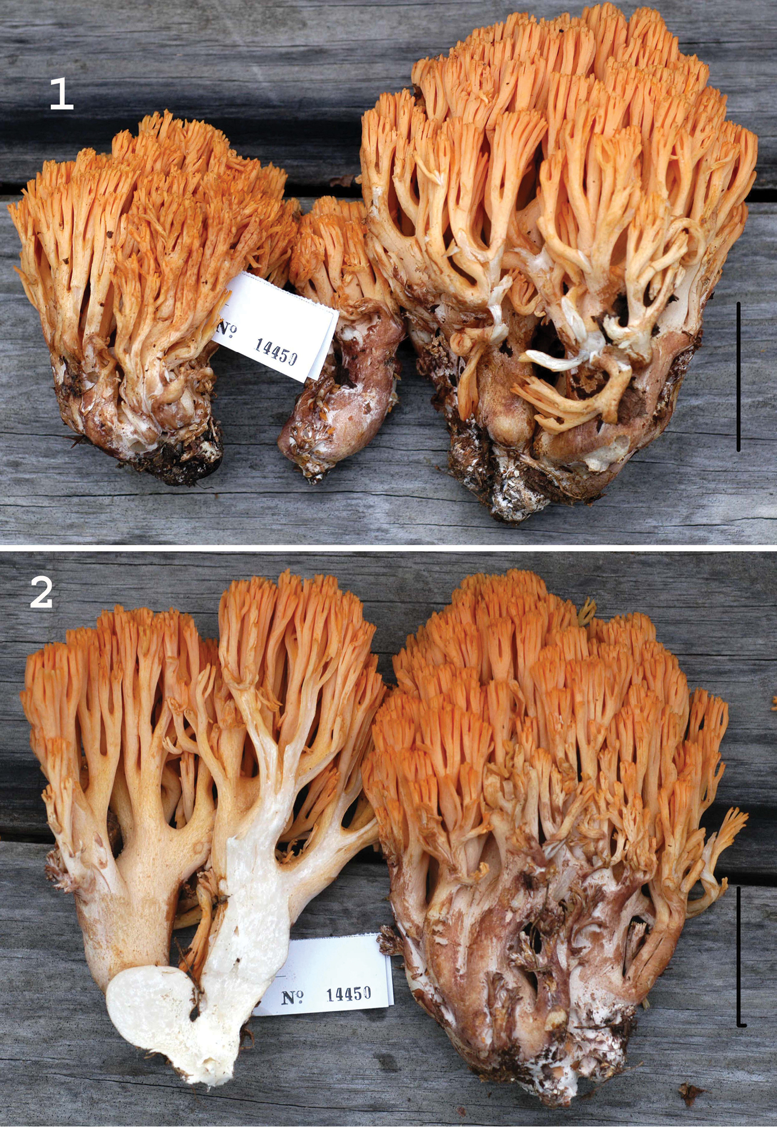
Basidiomata of Ramaria admiratia. 1 Exterior of two mature basidiomata and one immature 2 Left. Exposed stipe and lower branch trama. Right. Exterior of mature basidioma. Standard lines = 5 cm. Holotype.

Figures 3, 4.

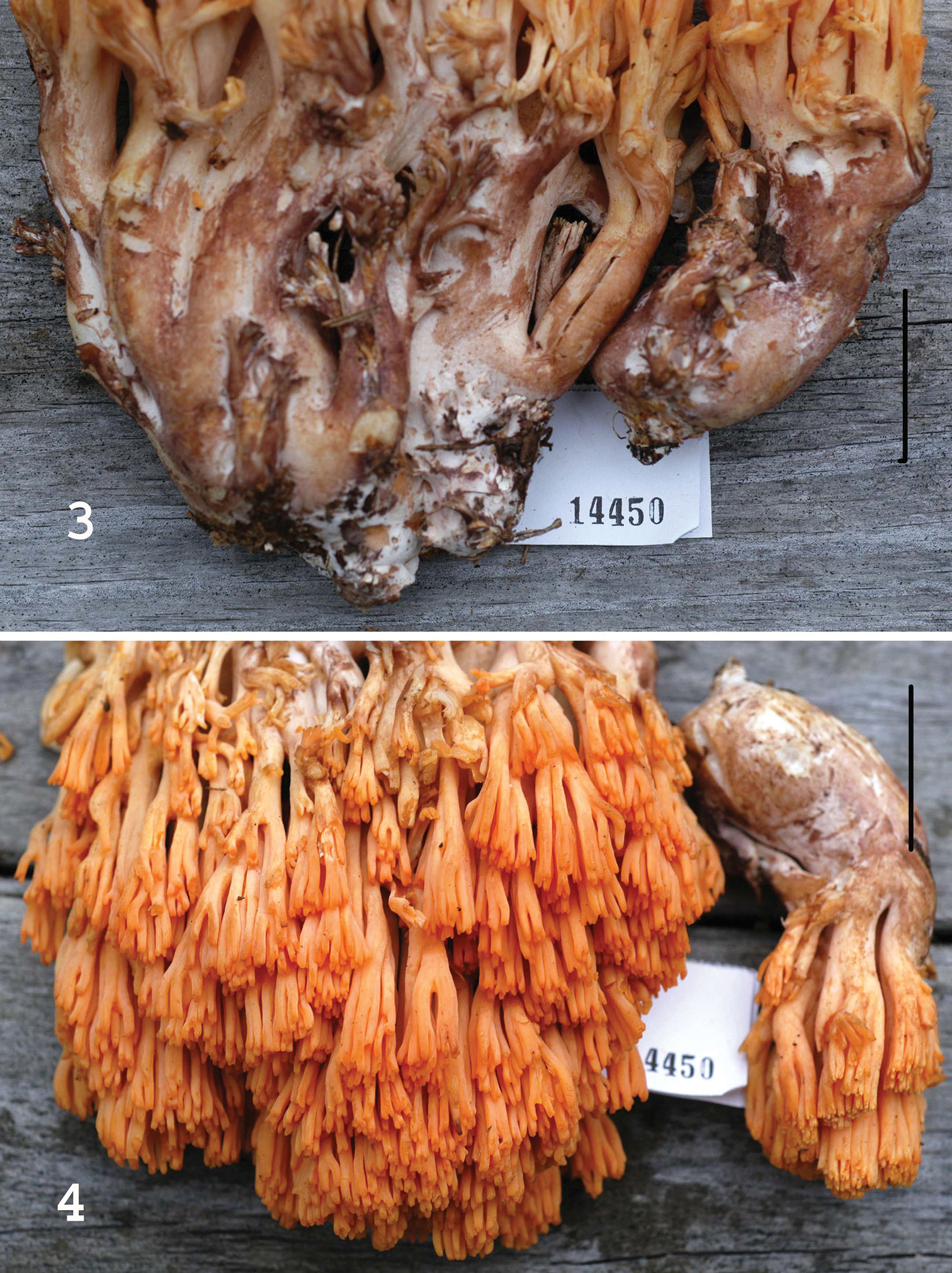
Basidiomata of Ramaria admiratia. 3 Stipe exteriors showing extensive brown bruising 4 Upper branches and branch apices. Standard lines = 5 cm. Holotype.

Habitat and phenology.

Generally second-growth hardwood forest of Quercus with scattered Carpinus, Carya and Acer; sole specimen from late autumn.

Surface of lower stipe covered with felty white tomentum; tomentum hyphae 3–4 μm diam, clampless, firm- to thick-walled (wall –0.5 μm thick), hyaline; acanthodendroid hyphae (Fig. 5B) common, refringent (PhC), strongly cyanophilous, 3–4 μm diam, thick-walled (wall –0.7 μm thick), appearing thorny. Hyphal construction of stipe medulla monomitic (with gloeoplerous hyphae), liberating significant debris in squash mounts; hyphae 4–14 μm diam, firm- to thick-walled (wall –0.5 μm thick), hyaline, clampless; ampulliform septa 7–17 μm diam, ampulliform to subspherical, thick-walled (wall –1 μm thick), delicately ornamented; gloeoplerous system represented by short lengths of cyanophilous hyphae with occasionally swellings. Hyphae of upper branch trama strictly parallel, tightly packed, thin-walled, clampless; cells filamentous to keg-shaped; occasionally slender (3–4 μm diam), non-refringent hyphae meandering through trama. Gloeoplerous system represent by occasional short lengths of strongly cyanophilous hyphae without septa. Basidia (Fig. 5A) 45–57 × 11–13 μm, clavate (not significantly bulbous), clampless, 4-sterigmate; contents multiguttulate (guttules refringent, apparently scattered throughout; PhC). Basidioles filamentous, digitate, uninformative. Basidiospores (Fig. 5B) 12–14.5 × 4.5–5.5(–6) μm (Q = 2.27–2.80; Qm = 2.52; Lm = 13.20 μm), ellipsoid, distinctly roughened in profile but ornamentation indistinct (PhC); contents heterogeneous with one or more non-refringent vacuoles (vacuoles amorphous, appearing empty on slightly darker background; PhC); ornamentation moderately cyanophilous, of narrow, axially oriented, meandering low ridges, appearing scattered, small and low in profile.

Figure 5.

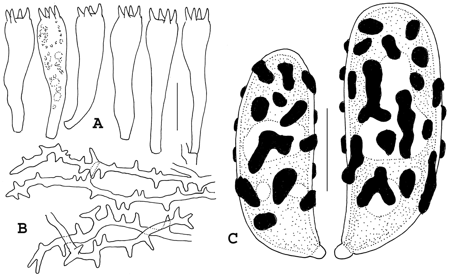
Microstructures of Ramaria admiratia. A Basidia B Acanthodendroidal hyphae of outer stipe tissue C Basidiospores showing cyanophilous ornamentation. Standard line for A, B = 20 µm; for C = 5 µm. Holotype.

Commentary.

Superficially, basidiomata of Ramaria admiratia resemble those of Ramaria cokeri through sordid yellow branches with reddish apices and brunnescent stipe. Ramaria cokeri belongs in Phaeoclavulina (Giachini 2004; Giachini and Castellano 2011; Giachini et al. 2001, 2010 = Ramaria subg. Echinoramaria) with echinulate basidiospores, clamped tramal hyphae and basidia, and rubribrunnescent (not brunnescent) stipe surface.

As noted by Christan (2008) taxa with cyanophilous dendrohyphidioid stipe tomentum hyphae are more common than originally envisioned (Marr and Stuntz 1973). Most common among them are taxa with yellow branches and apices and rather small stipes. In taxa resembling Ramaria admiratia, centering around Ramaria cystidiophora, varieties have been described based largely on odors (vars. anisata, fabiolens) and rubribrunnescent stipe (var. maculans) (see Marr and Stuntz 1973; Exeter et al. 2006). Conversely, Ramaria admiratia basidiomata exhibit a large, strongly brunnescent stipe and orange to red-orange branches and apices.

The terms “monomitic, ” “dimitic”, etc. were coined by E.J.H. Corner (1932) to describe the anatomy of various polypore basidiomata and have proven useful in subsequent, widely used taxonomic schemes. Dimitic referred only to presence of two hyphal types, specifically generative and skeletal, definition of the latter being philosophically augmented over some decades. Donk (1971) and Petersen (1975), conversely, while accepting the anatomical observations, linguistically considered dimitic to describe any tissue in which two hyphal types were found, whether generative and skeletal, generative and gloeoplerous, generative and acanthanophysoid, etc., as long as the hyphal types were accurately described. In the case of Ramaria admiratia (and other fleshy fungi with various second hyphal types), one is confronted with Cornerian terminology in which “monomitic” actually includes two hyphal types: generative plus cyanophilous, characteristically branched acanthophysoid hyphae.

Ramaria calvodistalis R.H. Petersen, sp. nov.

Mycobank no. 807955

Figs 6, 7
Holotype.

United States, Arkansas, Baxter Co., vic. Big Flat, Rte 341, Moccasin Creek Trailhead, Ozark National Forest, 36°02'N, 92°21'W, 24.X.2013, coll. RHP, TFB 14431 (TENN 69095).

Etymology.

Calvus = bald; distalis = referring to the spore wall opposite the hilar appendage.

Diagnosis.

1) Member of Ramaria subg. Laeticolora; 2) clamp connections absent from all tissues; 3) acanthodendroid hyphae absent; 4) stipe small, pruinose, white, without color change where handled or rubbed; 5) branches and apices yellow; 6) type locality northern Arkansas; 7) ITS sequence unique in the subgenus; (GenBank accession KJ416132).

Adult basidiomata (Fig. 6) –15 × 12 cm, repeatedly branched, coralloid; young basidioma with discrete base, white, hardly canescent or pruinose; adult basidiome base falsely fasciculate (i.e. discrete but with narrow grooves and crevices giving the appearance of several stipes strongly compressed), snow white, finely canescent where free of soil particles; abortive branchlets common, white; stipe flesh white, solid, firm, gelatinous only in areas of degeneration or maggot-infestation, without brown bands or patches; lower branches “orange buff” (5A5), upward becoming “warm buff” (5A4) to “antimony yellow” (4B6); apices rounded, concolorous. Odor none. Taste none; consistency mealy. No bruising reactions on surface or flesh.

Figure 6.

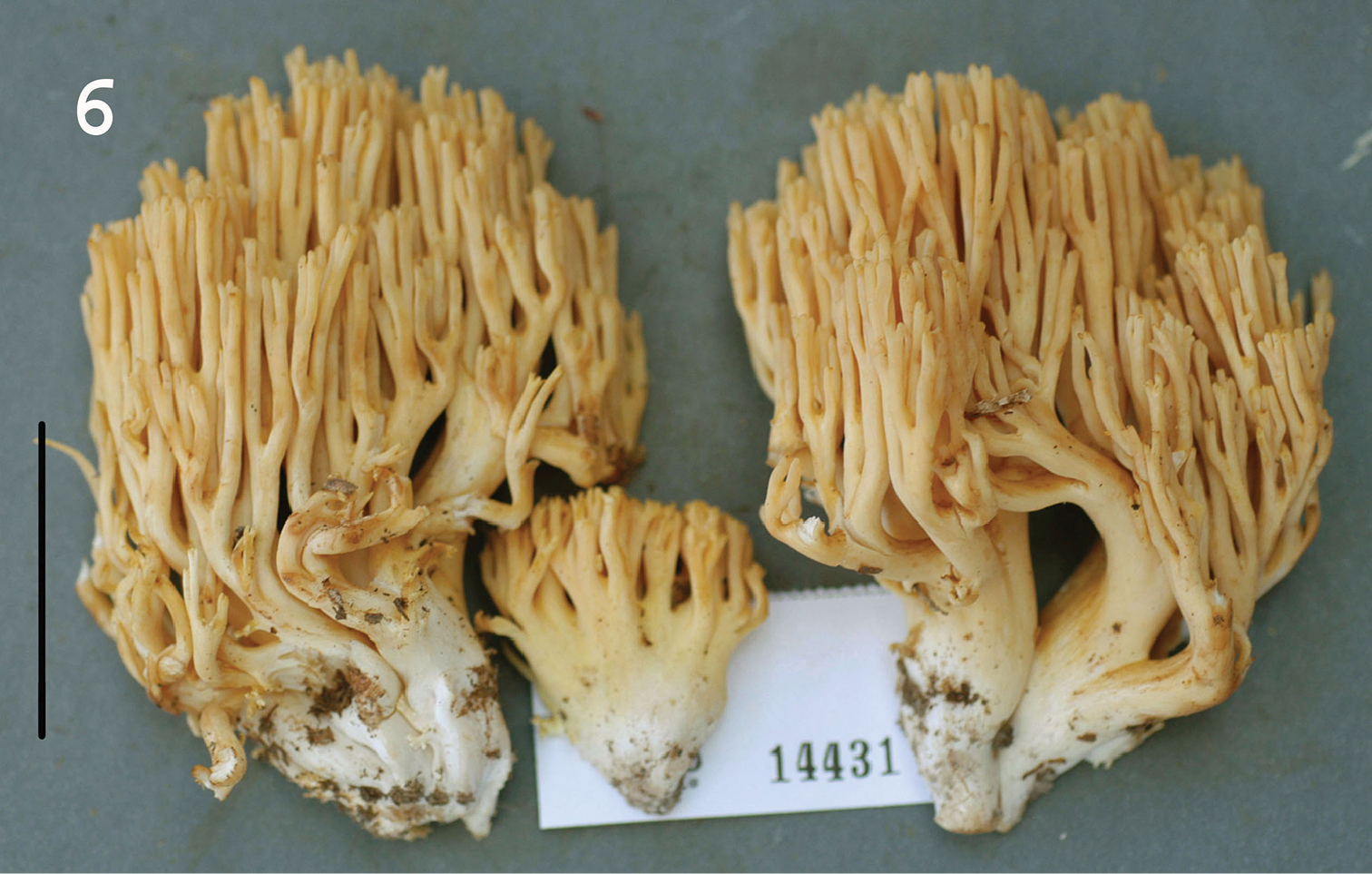
Basidiomata of Ramaria calvodistalis. Standard line = 5 cm. Holotype.

Habitat and phenology.

Possibly associated with deciduous trees from local forests of Quercus, Carya, Carpinus and occasional Pinus, solitary to gregarious, often in troops or rings; fruiting in late autumn.

Hyphae of stipe canescence 2–4 μm diam, relatively brittle and straight, firm-walled, rarely septate, without clamp connections, non-refringent; acanthodendroid hyphae absent; in non-gelatinous areas of stipe flesh hyphae 3–12 μm diam, tortuous, frequently branched, thick-walled (wall –0.7 μm thick), often refringent (PhC), without clamp connections; rare ampulliform swellings (without clamp connection) –16 μm diam, delicately ornamented internally, not unusually thick-walled. Hyphae of upper branch trama appearing subgelatinous under low magnification, but when squashed in KOH shown to be free, 3.5–12 μm diam, without clamp connections, firm-walled (wall –0.5 μm thick); cells filamentous to elongate-barrel-shaped. Basidioles often misshapen, paraphysoid, with various small lobes or sinuate shapes. Basidia (Fig. 7A) 55–72 × 12–13 μm, clavate with somewhat bulbous apex, 4-sterigmate, occasionally with an asymmetric lobe, without clamp connections; contents usually with proximal and distal refringent guttules. Basidiospores (Fig. 7B) (12–)14–15 × 4.5–5.0(–5.5) μm (Q = 2.67–3.33; Qm = 3.03; Lm = 14.05 μm), generally boletoid, with scattered small cyanophilous warts and patches through midsection of the spore but absent from the distal end which appears bald; contents with amorphous deposits (PhC); wall slightly thickened through midsection (wall –0.5 μm thick).

Figure 7.

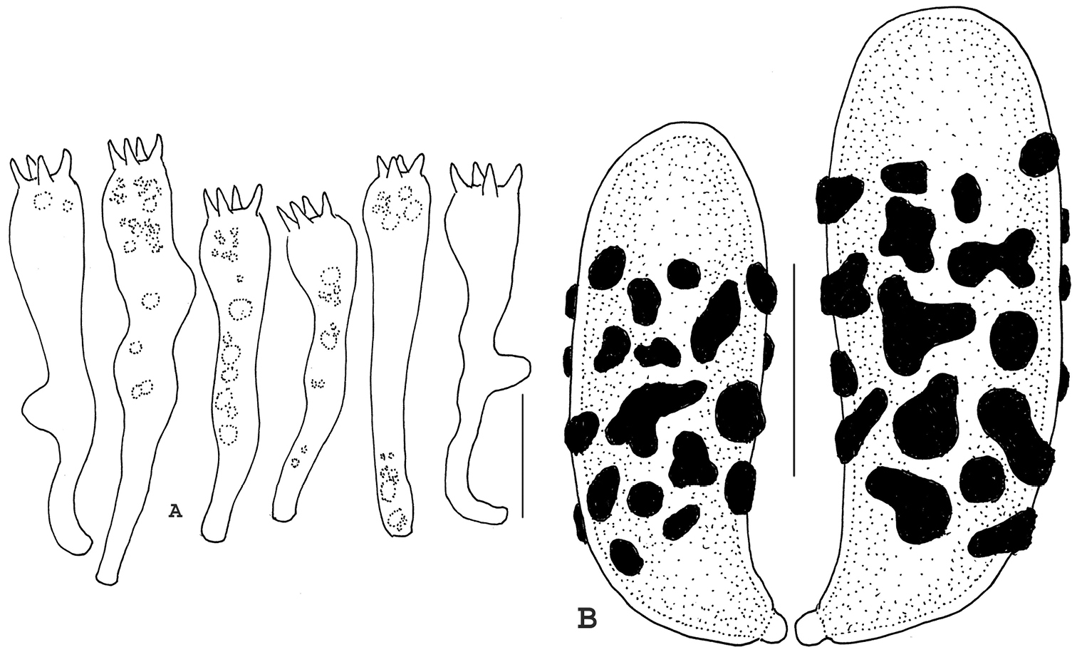
Basidia and basidiospores of Ramaria calvodistalis. A Basidia B Basidiospores. Standard line for A = 20 µm; for B = 5 µm. Holotype.

Commentary.

Care must be taken to ascertain the condition of basidial septa. In clamped basidia, subsequent basidioles arise through the subtending clamp. In clampless basidia (as above), subsequent basidioles arise in precisely the same fashion, arising just below the subtending septum, but without the telltale evidence of a clamp connection. Additional care is required to ascertain that both tramal hyphae and basidia are without clamps. Spores are among the longest in the subgenus and largely as a result, Qm value is high. Spore outline is distinctly boletoid with slight suprahilar depression. First impression was of delicately marbled stipe flesh (i.e. with small, scattered areas of hyphae with gelatinized walls), but closer examination revealed scattered degeneration of inner stipe flesh through maggot infestation. Lower and middle branches as well as apices are essentially unicolorous, straw yellow or dull ochraceous yellow. Juvenile branches exhibit a slight blush of pale pinkish buff, but this soon fades and slowly turns to the adult yellow shades.

In the three days of the NAMA foray, numerous collections of this species were made. Basidiomata seem to occur in troops and “fairy rings” under deciduous trees and are often somewhat bulky. Stipes are not deeply rooted and are easily dislodged, but adult stipes seem consistently maggot-ridden. Because a new taxon was not anticipated, only the type collection was preserved. Although the literature dealing with Ramaria of the Pacific Northwest has been summarized at least twice over the decades (Marr and Stuntz 1973; Exeter et al. 2006), modern literature is unavailable for Ramaria east of the Rocky Mountains except in fragments. Even less adequate is coverage of central United States, including the Ozark Mountains of Arkansas.

Marr and Stuntz (1973) described a small group of Ramaria taxa which exhibited cyanophilous “acantho-dendroid” hyphae in the outer stipe flesh, using Ramaria cystidiophora as the focal taxon, but including several infraspecific taxa. In all cases, stipes are white and pruinose and all upper basidiome parts are some shade of yellow. Exeter et al. (2006) illustrated basidiomata of several of the infraspecific taxa in the Ramaria cystidiophora complex, which macroscopically are reminiscent of Ramaria calvodistalis, so a special search was made for acanthodendroidal hyphae. Not only were no such hyphae found, but Ramaria calvodistalis lacks clamp connections, also a violation of the Ramaria cystidiophora complex.

If the key to clampless taxa in Exeter et al. (2006) is employed, no adequate match is found. Ramaria longispora produces spores of appropriate dimensions, but while upper branches and apices are yellow, lower branches are cantaloupe or pinkish salmon. In the working key of one of us (RHP), no match is found because the combination of yellow basidiome coloration, lack of clamp connections and long spores eliminates all candidates.

Ramaria admiratia and Ramaria calvodistalis LSU sequences place them near sequences representing brightly colored Ramaria species (Ramaria aurantiisiccescens and Ramaria araiospora) in subgenus Laeticolora (Fig. 8). ITS divergence within this subgenus is large, however, and Ramaria admiratia and Ramaria calvodistalis ITS sequences are only 86% similar to each other. Ramaria calvodistalis ITS sequences are most closely related (>97%) to two unnamed collections from Mexico (GenBank KC152173 and KC152176). These three collections differ from each other predominantly in the number of bases in repeat areas and probably represent the same lineage. We have previously noted that Mexico may have served as a glacial refugium for taxa now found further north (Hughes et al. 1999; Lickey et al. 2002; Hughes and Petersen 2004). There are no close blast matches to Ramaria admiratia in GenBank or in our sequence data set. A number of taxa in Fig. 8, based on assigned names, appear to be polyphyletic suggesting that morphological species concepts harbor more than one cryptic species or that misidentifications are common.

Figure 8.

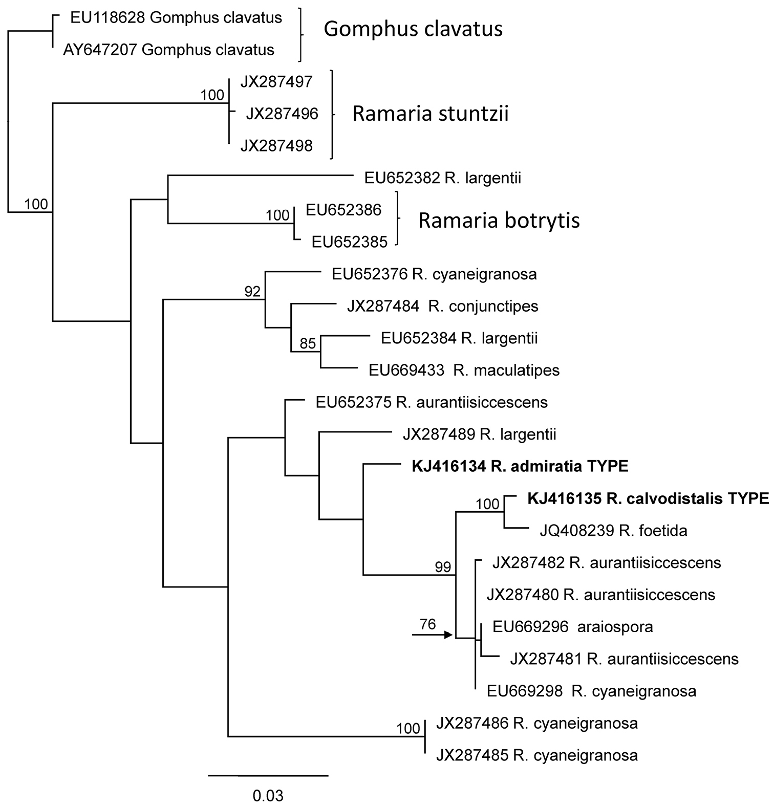
PhyML Phylogeny of proposed new species of Ramaria based on nrLSU sequences. Bootstrap support equal or greater than 70% is given to the left of the supported node. GenBank accession numbers are given at the end of each twig.

Acknowledgements

Sincere thanks are extended to Carl Davis and Therese Martin of Louisiana who collected Ramaria admiratia. Work was partially supported by NSF DEB1144974 to RHP and KWH.

References
Burt EA (1922) The North American species of Clavaria. Annals of the Missouri Botanical Garden 9: 1–78. doi: 10.2307/2989963, http://www.jstor.org/stable/2989963
Christan J (2008) Die Gattung Ramaria in Deutchland. IHW Verlag, 352 pp.
Christan J, Hahn C (2005) Zur Systematik der Gattung Ramaria (Basidiomycota, Gomphales). Zeitschrift für Mykologie.
Coker WC (1923) The Clavarias of the United States and Canada. University of North Carolina Press, 209 pp. http://www.cabdirect.org/abstracts/19241100863.html;jsessionid=153D33A681E03C255882FF93A578684E?freeview=true
Corner EJH (1932) The fruit-body of Polystictus xanthopus. Annals of Botany 46: 71–111.
Corner EJH (1950) A monograph of Clavaria and allied genera. Annals of Botany Memoirs Series 1: 1–740.
Corner EJH (1970) Supplement to A monograph of Clavaria and allied genera. Nova Hedwigia, Beiheft 33: 1–299.
Donk MA (1971) Multiple convergence in the polyporaceous fungi. In: Petersen RH (Ed) Evolution in the higher Basidiomycetes. University of Tennessee Press, Knoxville, 393–422.
Doty MS (1944) Clavaria, the species known from Oregon and the Pacific North West. MS Thesis, Oregon St. Coll., Corvallis, 91 pp.
Exeter RL (2001) Ramaria of the Pacific Northwest. Bureau of Land Management, Salem, OR, 40 pp.
Exeter RL, Norvell L, Cázares E (2006) Ramaria of the Pacific Northwestern United States. Bureau of Land Management, Salem, OR, 157 pp.
Franchi P, Marchetti M (2001) Introduzoione allo studio dei funghi clavarioidi. 84th incontro del Centro Micologico Lombardo, Monte Barro 20-05-2001. Corso di aggriorbamento per micologi 21: 1–69.
GCG (2000) Genetics Cooperative Group Wisconsin Package, Version 10.3. Accelrys Inc., San Diego, California.
Geneious (2005) Geneious version 6.1.6 created by Biomatters. http://www.geneious.com/
Giachini AJ (2004) Systematics of the Gomphales. The genus Gomphus Pers. sensu lato. PhD Thesis, Oregon State University, Corvallis, Oregon.
Giachini AJ, Castellano MA (2011) A new taxonomic classification for species in Gomphus sensu lato. Mycotaxon 115: 183–201. doi: 10.5248/115.183
Giachini AJ, Spatafora JW, Cázares E, Trappe JM (2001) Molecular phylogenetics of Gomphus and related genera inferred from nuclear large and mitochondrial small subunits ribosomal DNA sequences. Abstract, III International Conference of Mycology, Adelaide, Australia, 95.
Giachini AJ, Hosaka K, Nouhra ER, Spatafora JW, Trappe JM (2010) Phylogenetic relationships of the Gomphales based on nuc-25S-rDNA, mit-12S-rDNA and mit-ATP6-DNA combined sequences. Fungal Biology 114: 224–234. doi: 10.1016/j.funbio.2010.01.002
Hughes KW, McGhee LL, Methven AS, Johnson J, Petersen RH (1999) Patterns of geographic speciation in the genus Flammulina based on sequences of the ribosomal ITS1-5.8S-ITS2 area. Mycologia 91: 978–986. doi: 10.2307/3761628
Hughes KW, Petersen RH (2004) Origins of Fungal Biodiversity in the Southern Appalachians. Inoculum 55(4): 18. [abstract]
Hughes KW, Petersen RH, Lodge DJ, Bergemann S, Baumgartner K, Tulloss RT, Lickey EB, Cifuentes Blanco J (2013) Evolutionary consequences of putative intra- and interspecific hybridization in agaric fungi. Mycologia 105: 1577–1594. doi: 10.3852/13-041
Humpert AJ, Muench EL, Giachini AJ, Castellano MA, Spatafora JW (2001) Molecular phylogenetics of Ramaria and related genera: evidence from nuclear large subunit and mitochondrial small subunit rDNA sequences. Mycologia 93: 465–477. http://www.jstor.org/stable/3761733
Kornerup A, Wanscher JH (1967) Methuen handbook of colour. 2nd revised edition. Methuen Co., London, 243 pp.
Lickey EB, Hughes KW, Petersen RH (2002) Biogeographical patterns in Artomyces pyxidatus. Mycologia 94: 461–471. doi: 10.2307/3761780
Marr CD, Stuntz DE (1973) Ramaria of western Washington. Bibliotheca Mycologica 38: 1–232.
Petersen RH (1975) Ramaria subgenus Lentoramaria with emphasis on North American taxa. Bibliotheca Mycologica 43: 1–161.
Pine EM, Hibbett DS, Donoghue MJ (1999) Phylogenetic relationships of cantharelloid and clavarioid Homobasidiomycetes based on mitochondrial and nuclear rDNA sequences. Mycologia 91: 944–963. doi: 10.2307/3761626
Rambaut A (2006) FigTree: Tree Figure Drawing Tool Version 1.4.0 2006–2012, Institute of Evolutionary Biology, University of Edinburgh. http://tree.bio.ed.ac.uk/software/figtree/
Ridgway R (1912) Color standards and color nomenclature. Publ. Priv., Washington, DC. [53 figs]
Schild E (1995) Eine Analyse de Ramaria-fennica-fumigata-versatilis-Komplexes. Zeitschrift für Mykologie 61: 123–130.