


(C) 2012 Michel N. Benatti. This is an open access article distributed under the terms of the Creative Commons Attribution License 3.0 (CC-BY), which permits unrestricted use, distribution, and reproduction in any medium, provided the original author and source are credited.
For reference, use of the paginated PDF or printed version of this article is recommended.
Descriptions are presented for the seven known Bulbothrix (Parmeliaceae, Lichenized Fungi) species with salazinic acid in the medulla and without vegetative propagules. Bulbothrix continua, previously considered as a synonym of Bulbothrix hypocraea, is recognized as independent species. The current delimitations are confirmed for Bulbothrix enormis, Bulbothrix hypocraea, Bulbothrix meizospora, Bulbothrix linteolocarpa, Bulbothrix sensibilis, and Bulbothrix setschwanensis. New characteriscs and range extensions are provided.
Parmeliaceae, Parmelinella, norstictic acid, bulbate cilia
The genus Bulbothrix Hale was proposed for the group of species called Parmelia Series Bicornutae (Lynge) Hale & Kurokawa (
During an unpublished revision of the genus Bulbothrix (
For a comprehensive understanding and easy assessment of all the data on the review of this genus comprising ca. 60 species gathered in an unpublished review study by
The descriptions of the species treated here can also be found somewhere else in the literature such as
Type material and additional specimens were studied from BM, FH, GLAM, H, HUFSCAr, LD, LG, M, NY, S, SP, TNS, TUR, US, W, and WU, originating from Asia, Africa, and South America. Added is a considerable material collected in Brazil during the last 30 years, mainly by the author and the members of the Lichenological Study Group of the Instituto de Botânica (GEL) in Brazil.
The methodology and conventions are detailed in
The presence of salazinic acid is indicated by a K+yellow→dark red spot test reaction, not unlike that of norstictic acid, but turning darker red even with different KOH concentrations (10% and 30%) in Bulbothrix specimens. It also reacts P+ yellow, and does not react to C or KC, neither reacts to UV light. Its presence can also be indicated by the formation of bundles of thin elongated crystals of a deep reddish color, visible under a light microscope after the transfer of a small piece of the thallus or of the apothecia onto a microscope slide and dropping the reagent on the fungal material. However, as compared with the much more obvious crystals of norstictic acid (
The species selected for comparisons are those who show close morphological or chemical similarities, and those most often compared by other authors due to peculiar characteristics (e.g.,
The study confirmed all seven previously known species containing salazinic acid that do not form vegetative propagules or pustules. Four species, Bulbothrix continua, Bulbothrix linteolocarpa, Bulbothrix sensibilis and Bulbothrix setschwanensis are corticolous, while Bulbothrix enormis is saxicolous. Bulbothrix hypocraea and Bulbothrix meizospora are predominatly corticolous, rarely saxicolous and in the case of Bulbothrix meizospora, also rarely terricolous. All species are described in detail and discussed below.
Table 1 summarizes the main characteristics (usual averages found) used for differentiate the species in this paper and most commonly accepted in literature (see e.g. list in the Introduction).
Comparative diagnostic characteristics of Bulbothrix species containing salazinic acid that do not reproduce by vegetative propagules. The data refer to the most typical range found.
| Species | Laciniae width | Maculae | Marginal bulb size | Lower cortex color | Ascospore size |
|---|---|---|---|---|---|
| Bulbothrix continua | 1–2.5 mm | absent | ca. 0.05–0.15 mm wide |
brown to pale brown |
9.0–13.5 × 5.0–7.5 µm |
| Bulbothrix enormis | 1.5–8 mm | absent | ca. 0.05–0.20 mm wide |
mostly pale brown |
7.0–11.5 × 5.0–7.0 mm |
| Bulbothrix hypocraea | 1–2.5 mm | present (abundant) |
ca. 0.10–0.30 mm wide |
pale brown to ivory |
7.0–14.0 × 5.0–8.0 mm |
| Bulbothrix linteolocarpa | < 1 mm | absent | ca. 0.05–0.10 mm wide |
pale brown | 9.0−16.0 × 6.5−8.0 µm |
| Bulbothrix meizospora | 1.5–6 mm | present (weak) |
ca. 0.10–0.30 mm wide |
black center and most margins | 10.0−22.0 × 7.5−14.0 µm |
| Bulbothrix sensibilis | 1.5–5 mm | present (variable) |
ca. 0.05−0.25 mm wide |
black center with brown margins | 7.0−13.0 × 5.0−7.0 µm |
| Bulbothrix setschwanensis | 1–5 mm | absent | ca. 0.05−0.25 mm wide |
pale brown center and margins | 10.0−19.0 × 6.0−10.0 µm |
Mycobank: MB 341595
Figures 1–2Brasiliae civit Matto Grosso, Serra da Chapada, Buriti, leg. Malme s.n., 19-VI-1894 (S!).
Thallus subirregularly laciniate, grayish green in the herbarium, up to 3.8 cm diam., subcoriaceous, corticolous; upper cortex 15.0−22.5 µm thick, algal layer 25.0−37.5 µm thick, medulla 67.5−85.0 µm thick, lower cortex 20.0−25.0 µm thick. Laciniae anisotomically to irregularly dichotomously branched, 0.8–1.9 (-2.3) mm wide, slightly imbricate, rarely becoming crowded at the center, adnate and adpressed, with flat, subtruncate apices; margins plane, smooth and sinuous to crenate, entire, occasionally sublacinulate; axils oval. Upper surface smooth and continuous, becoming rugose and irregularly cracked in some parts; laminal ciliary bulbs absent. Adventitious marginal lacinulae scarce and restrict to older parts, short, 0.2–0.5 × 0.1–0.5 mm, plane, simple to rarely furcate; apices truncate; lower side concolorous to the lower marginal zone. Maculae absent. Cilia black, without or with simple apices, commonly bent downwards, 0.05–0.40 × ca. 0.03 mm, with semi-immerse to emerse bulbate bases 0.05–0.15 (-0.25) mm wide, abundant throughout the margin spaced 0.05−0.10 mm from each other to contiguous, solitary or in small groups at the crenae and axils, scarce at the apices of the laciniae. Soredia, Pustulae and Isidia absent. Medulla white. Lower surface brown to pale brown, shiny to opaque, smooth to subrugose, weakly papillate, moderately rhizinate. Marginal zone brown to pale brown, indistinct from the center, shiny to opaque, smooth, weakly papillate, weakly to densely rhizinate. Rhizinae black to pale brown brown, partially white or with whitish apices when close to the margins, simple or sometimes irregularly branched, commonly with bulbate bases, 0.10–0.65 × 0.03–0.10 mm, frequent but becoming abundant close to the margins or scarce at some other parts, sometimes agglutinated, evenly distributed. Apothecia concave to plane or convex, adnate to sessile and distended over the laciniae, 0.4–3.7 mm diam., laminal; margin smooth to subcrenate, ecoronate; amphithecium smooth, without ornamentations. Disc pale brown, epruinose, imperforate; epithecium 15.0–20.0 mm high; hymenium 50.0−62.5 µm high; subhymenium 15.0−22.5 µm high. Ascospores ellipsoid to oval, 9.0–13.5 × 5.0–7.5 µm; epispore ca. 1.0 mm. Pycnidia common, laminal, immersed, with black ostioles. Conidia baciliform 5.0−7.5 × 1.0 µm.
TLC/HPLC: cortical atranorin, medullary salazinic and consalazinic acids (see also Hale 1976).
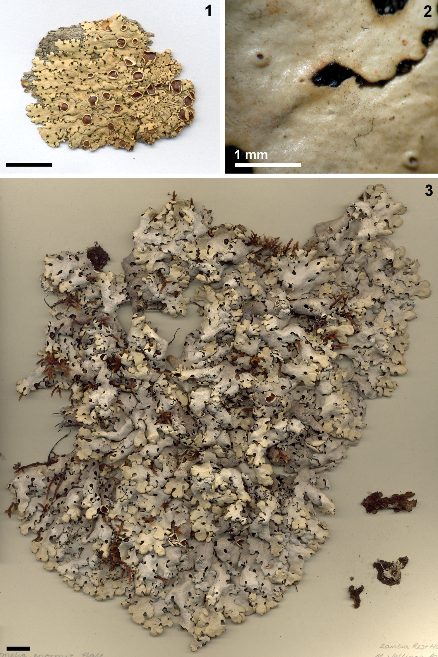
1 Holotype of Bulbothrix continua 2 Detail of the shiny emaculate upper cortex 3 Holotype of Bulbothrix enormis. Scale bars = 1 cm (1, 3), 1 mm (2).
South America: Brazil: State of Mato Grosso (
Brazil, Mato Grosso State, Santa Anna da Chapada, Buriti Municipality, leg. G. O. Malme s.n., 19-IV-1894 (US). Idem, São Paulo State, 6 km SW of Jaboticabal, 21°35'S, 48°35'W, on trees in “cerradão” (savannah), leg. A. Fletcher 10108, 1-V-1975 (BM). Idem, São Manuel Municipality, Fazenda Palmeira da Serra, unofficial private cerrado (savannah) reserve, on tree trunk in the cerrado, leg. M. P. Marcelli & S. B. Barbosa 35232, 03-VI-2003 (SP, paratype of Bulbothrix vainioi). Idem, Santa Rita do Passa Quatro Municipality, Fazenda Vassununga, km 259 of Anhanguera Highway, 760 m, transition from cerrado to “cerradão” (savannah), trees with signs of old burns, on a tree thin twig, leg. M. P. Marcelli & S. B. L. Morretes 16055, 21-IX-1978 (SP). Idem, Moji-Guaçu Municipality, Fazenda Campininha, Estação Biológica de Moji-Guaçu, illuminated, dry savannah, on tree thin twig, leg. M. P. Marcelli 15885, 29-VI-1979 (SP).
The holotype (Figs 1–2) consists of a small, entire thallus, in good condition. The material contains several apothecia at different stages of maturity with well developed ascospores, and some pycnidia. It is on a small piece of tree bark but with the laciniae apices free from the substrate, and is not glued to cardboard. There is no trace of true maculae in the upper cortex or in the amphithecia, although the cortex is in fact somewhat pale and shiny.
Shortly after the recombination of ‘Parmelia’ continua into Bulbothrix (
Compared with the specimens of Bulbothrix continua, those of Bulbothrix hypocraea are always quite maculate, and their thalli often form wider laciniae than those of Bulbothrix continua.
Bulbothrix linteolocarpa Marcelli differs by the much narrower laciniae, barely exceeding 0.5 mm, that are also more linear with contiguous cilia forming long apices. As they mature, apothecia of Bulbothrix linteolocarpa continually adapt to the conformation of the surface, settling on the laciniae as if they were spreading over them. Two specimens of uncertain identity cited by
Bulbothrix sensibilis (Steiner & Zahlbruckner) Hale differs from Bulbothrix continua equally by the presence of cortical maculae and moreover by the shiny black lower cortex with dark brown margins. Bulbothrix setschwanensis (Zahlbruckner) Hale differs by the larger laciniae (ca. 1.5−5.0 mm wide) and by the size of the ascospores (usually 12.0−19.0 × 7.0−10.0 µm).
Mycobank: MB 360209
Figure 3Zambia, Zambia Rest House area, Nyika Plateau, 7600 ft., on granite rocks, M. Jellicoe s.n., VII-1968 (BM!, isotypes at TNS n.v. and US!).
Thallus sublinearly to subirregularly sublaciniate, gray with dusky green distal parts in herbarium, up to 24.1 cm diam., coriaceous, saxicolous; upper cortex 15.0−22.5 µm thick, algal layer 52.5−80.0 µm thick, medulla 120.0−150.0 µm thick, lower cortex 15.0−25.0 µm thick. Laciniae isotomically or anisotomically to irregularly dichotomously branched, (1.3−) 3.2–6.0 (−7.8) mm wide, imbricate to crowded, slightly to not adnate and loose, occasionally almost subcanaliculate, with involute to revolute or sometimes plane, subrounded to subtruncate apices; margins plane to subundulate or slightly involute, smooth and sinuous to occasionally subcrenate, entire, rarely little sublacinulate; axils oval. Upper surface smooth and continuous, rarely with some random irregular cracks; laminal ciliary bulbs absent. Adventitious marginal lacinulae scarce on random parts, short, 0.5–1.7 × 0.3–1.1 mm, usually involute, simple or sometimes irregularly branched; apices truncate to subtruncate; lower side concolorous to the lower marginal zone. Maculae absent. Cilia black to dark brown, with simple to partially double or furcate apices, occasionally bent downwards, 0.10–1.20 (-1.80) × ca. 0.05 (−0.10) mm, with semi-immerse to emerse bulbate bases 0.05–0.20 (-0.35) mm wide or partially not bulbate, sometimes disposed on a distinct black line, frequent to abundant throughout the margins, in small groups in the axils and adjacent parts spaced 0.10−0.40 mm from each other, becoming absent or scarce at the apices of the laciniae and adjacent parts. Soredia, Isidia and Pustulae absent. Medulla usually white, but pinkish in some random parts and below the hymenial discs. Lower surface pale brown, occasionally with random small dark brown or black spots, shiny, smooth, moderate to densely rhizinate. Marginal zone brown, indistinct from the center or sometimes interrupted by blackish spots, shiny, smooth, weakly papillate, gradually becoming rhizinate following the center. Rhizinae black to variably brown, occasionally whitish or with withish apices, simple to occasionally furcate or irregularly branched, without bulbate bases or with subtle basal or displaced bulbs, 0.20–1.80 (−2.30) × 0.05–0.10 mm, frequent to abundant, evenly distributed. Apothecia subconcave to urceolate or occasionally plane, becoming folded when old, adnate to substipiate, 1.1–10.0 mm diam., laminal to submarginal, ecoronate; margin smooth to subcrenate and fissured; amphithecium smooth, without ornamentations. Disc brown to dark brown, epruinose, imperforate; epithecium 7.5–17.5 µm high; hymenium 20.0−55.0 µm high; subhymenium 12.5−22.5 µm high. Ascospores ellipsoid to oval or subrounded, 7.0–11.5 × 5.0–7.0 mm; epispore (0.5−) 1.0−1.5 mm thick. Pycnidia laminal to submarginal, frequent, immerse, with brown or black ostioles. Conidia baciliform to weakly or distinct bifusiform 5.0−8.0 × 0.75 µm.
TLC/HPLC: cortical atranorin and chlororatranorin, medullary salazinic and consalazinic acids (see also
Africa: Zambia (
Zambia, Zambia Rest House area, Nyika Plateau, leg. M. Jellicoe s.n., IV-1969 (FH).
The holotype consists of a large specimen more than 20 cm in diameter, glued to board, in excellent condition and containing several apothecia and pycnidia. There are some loose fragments from 3 to 10 cm diam., allowing vizualization of the lower cortex details. The isotype from US consists of several loose fragments such as those with the holotype, also in good condition, with mature apothecia and pycnidia. There are no remains of the rocky substrate of where the materials were collected, indicating that the thalli were not strongly adhered to the substrate.
Originally,
Most of the cilia seen in the specimens studied are bulbate, but some of them are not, even including some of the largest cilia. However, the bulbs have the typical anatomical structure of Bulbothrix species, with an oily substance and idioblasts cells (
While
Bulbothrix hypocraea (Vainio) Hale and Bulbothrix setschwanensis (Zahlbruckner) Hale were compared to Bulbothrix enormis by
Bulbothrix haleana Sérusiaux (LG!, US!) differs by the thallus aspect, with narrow subirregular laciniae 1.0−3.5 mm wide, the overall globose and always evidently bulbate cilia with shorter apices, and by the smaller ascospores 5.0−9.0 × 4.0−7.0 µm. Further it contains norstictic acid, rather than salazinic acid as stated in the original description (
Another relatively similar species, Bulbothrix meizospora (Nylander) Hale, also differs by the narrower laciniae (ca. 1.5−4.0 mm wide), larger ascospores (12.5−22.0 × 9.0−14.0 µm) and a black lower cortex with brown margins.
Mycobank: MB 341600
Figures 4–9Angola, Huilla (3800 ad 5500 ped. s. m.), ad corticem arborum Leguminosarum in sylvis densis juxta flumen Monino, ca. 14°16°S, leg. Welwitsch 32 pro parte, IV-1860 (TUR-V!, duplicate at BM!).
Thallus sublinearly to subirregularly laciniate to sublaciniate, light dusky gray in the herbarium, in fragments up to 5.2 cm diam., coriaceous to subcoriaceous, corticolous or rarely saxicolous; upper cortex 15.0−25.0 µm thick, algal layer 25.0−42.5 µm thick, medulla 75.0−125.0 µm thick, lower cortex 12.5−20.0 µm thick. Laciniae anisotomicaly dichotomously to irregularly branched, (0.5−) 0.9–2.6 (−3.0) mm wide, contiguous to slightly imbricate, rarely crowded at the center, ±adnate and loosely adpressed, with plane to slightly involute or revolute, truncate, subtruncate or subrounded apices; margins plane to subplane, smooth to sinuous and subcrenate or subirregular, entire to slightly incised, not lacinulate; axils oval to irregular. Upper cortex mostly continuous, occasionally with some irregular cracks on older parts, smooth to subrugose; laminal ciliary bulbs absent. Adventitious marginal lacinulae absent, even on old parts. Maculae usually distinct, puntiform to efigurate, laminal on the thallus or on the amphithecia of the apothecia. Cilia black or rarely brown, without or with simple apices, often bent downwards, 0.05–0.65 × 0.03–0.05 mm, with semi-immerse to emerse, bulbate bases (0.05-) 0.10–0.30 mm wide (these partially enlarged or occasionally absent), frequent throughout the margins, solitary or in small groups in the crenae and axils spaced 0.05−0.20 mm from each other to occasionally contiguous, becoming absent or scarce at the apices of the laciniae and adjacent parts, usually absent or scarce in the apices of the laciniae and adjacent parts. Soredia, Isidia and Pustulae absent. Medulla white. Lower surface pale brown to ivory, opaque to slightly shiny, smooth, moderately rhizinate, sometimes up to the margins. Marginal zone indistinctly delimited from the center to slightly attenuate, 0.5–2.0 mm wide, pale brown to ivory, opaque to slightly shiny, smooth, weakly papillate, often rhizinate. Rhizinae ivory or light to dark brown, occasionally blackish, whitish or with white apices, simple or sometimes irregularly branched, partially with blackish bulbate bases or displaced bulbs, 0.10–0.80 (–1.10) × 0.05–0.10 mm, frequent, sometimes agglutinated, evenly distributed. Apothecia subconcave to subplane, becoming folded when old, sessile to adnate to substipiate, 0.3–8.2 mm diam., laminal to submarginal, ecoronate; margin subcrenate; amphithecia smooth occasionally fissured, without ornamentations. Disc pale brown to reddish brown, epruinose, imperforate; epithecium 7.5–17.5 µm high; hymenium 32.5−70.0 µm high; subhymenium 10.0−37.5 µm high. Ascospores ellipsoid to oval or subrounded, 7.0–14.0 × (5.0–) 6.0–8.0 mm; epispore ca. 1.0 mm. Pycnidia laminal, frequent mainly at the distal parts of the laciniae, immersed, with black ostioles. Conidia baciliform to weakly bifusiform (4.0−) 5.0−9.0 × 0.75 µm.
TLC/HPLC: cortical atranorin and chloroatranorin, medullary salazinic and consalazinic acids (see also Hale 1976).
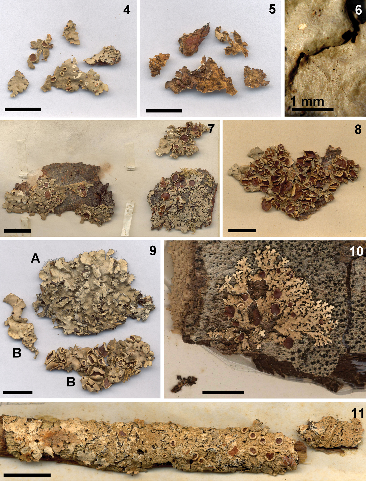
4 Lectotype of Bulbothrix hypocraea 5 Detail of the lower side of the lectotype 6 Detail of the maculate upper cortex 7 Duplicate of Bulbothrix hypocraea 8 Holotype of Parmelia leptascea 9 Lectotype of Parmelia proboscidea var. saxicola (marked B) 10 Holotype of Bulbothrix linteolocarpa 11 Holotype of Bulbothrix meizospora. Scale bars = 1 cm (4, 5, 7, 8, 9, 10, 11), and 1 mm (6).
Africa (
Africa, Bakoba am Victoriasee, auf Baumrinden, Schröder 319 (W!, holotype of Parmelia leptascea). Rhodesia (Zimbabwe), District Salisbury, Chindamora Reserve, Ngomakukira, epiphyte on Julbernardia globiflora, Swatzia madagascariensis etc., leg. H. Wild 5806, 10-VI-1962 (NY). Kenia, K4, Nth. Nyeri (1°30'S, 37°30'E), Lew Downs Ranch, 0 km W of Isiolo, Acacia woodland, leg. H. Ballev 660c, 4-XII-1981 (NY). Tanzania, Mahulo, Kipengere, loc. c. s. on rock, with Usnea densirostra, very mixed, leg. Eusébio 13 bis, 02-III-1935 (FI!, lectotype of Parmelia proboscidea var. saxicola, designate here as “B”). Brazil, Mato Grosso State, Serra do Roncador, riverine forest, 46 km north of Chavantina, Rio Vau, epiphyte, abundant, leg. G. T. Prance & N. T. Silva 59380A, 11-X-1964 (NY). Idem, Minas Gerais State, Lagoa Santa, leg. Warming s.n (M). Idem, São Paulo State, Brotas Municipality, NW side of intersectionof road to Campo Alegre with the Brotas-Itirapina road, arboreal semi-closed cerrado woodland, 22°17'S, 47°56'W, 750 m, leg. G. Eiten et al. 2976c, 16-VI-1961 (US). Idem, Santa Rita do Passa Quatro Municipality, fazenda Vassununga, km 259 of Anhanguera highway, on woody stem of vine, leg. M. P. Marcelli & B. L. Morretes 15628, 03-VI-1978 (SP). Idem, São Manuel Municipality, Fazenda Palmeira da Serra, non official particular cerrado (savannah) reserve, on tree trunk at the woods, leg. M. P. Marcelli & S. B. Barbosa 35680, 03-VI-2003 (SP).
The lectotype (Figs 4–5) consists of three small fragments on bark glued to cardboard, and some smaller fragments packed in paper, free of substrate. The duplicate (Fig. 7) consists of three fragments, all on bark, one of them glued to cardboard (fragments free from substrate were used to see the features of the lower cortex). The type material has several pycnidia, restricted to the distal parts of the laciniae.
Bulbothrix hypocraea has the most strongly maculate thalli of the genus (Fig. 6). However, in very old herbalium material, such as the type, the maculae may become difficult to be see due to the darkening of the upper cortex and the staining of the medulla by the oxidized salazinic acid. In this case, a bright illumination and wetting the thalli make the maculae more visible.
Most cilia have an evident bulbate base, their apices are usually bent downwards and sometimes barely visible from above. Some cilia, however, have no basal bulb, but just a thickened, tapering base (possibly an early stage in the development of the cavity).
The color of the lower cortex varies from brown to ivory or cream, the marginal zone being slightly darker than the center (Fig. 5). The ivory color is the least common, and is similar to that observed in the lower margin of other Parmeliaceae (like Parmotrema species) which are white ivory when fresh, eventually changing color after time in the herbarium.
The swellings seen in the rhizines along its length are not actually endociliary pycnidia, as first suspected by
The ascospore measurements provided by
The description by
The type collection of Parmelia proboscidea var. saxicola Cengia Sambo (FI!) consists of a ciliate Parmotrema specimen with submarginal, pustular soralia, and two fragments of Bulbothrix hypocraea (Fig. 9, marked B) that make up the majority of the collection. Therefore the latter are appointed here as the lectotype, as it is in accordance to the species protologue. The comments of Cengia
Bulbothrix setschwanensis (Zahlbruckner) Hale differs by the absence of cortical maculae and by larger ascospores 12.0−19.0 × 6.0−9.0 µm.
Bulbothrix linteolocarpa Marcelli was compared to Bulbothrix hypocraea by
Among other similar species, Bulbothrix sensibilis (Steiner & Zahlbruckner) Hale was compared to Bulbothrix hypocraea by
Mycobank: MB 458790
Figure 10Brazil, Mato Grosso State, between Jaciara and São Vicente, km 313 of BR-364 highway, ca. 100 km ESE of Cuiabá, cerradão (savannah), on tree trunk, leg. Marcelli 8446, 2-VII-1980 (SP!).
Thallus sublinear laciniate, dusky gray in the herbarium, up to 2.6 cm diam., subcoriaceous, corticolous; upper cortex 20.0−30.0 µm thick, algal layer 55.0−75.0 µm thick, medulla 25.0−35.5 µm thick, lower cortex 10.0−15.0 µm thick. Laciniae irregularly to anisotomically dichotomously branched, 0.2–0.6 (-0.8) mm wide, contiguous to occasionally slightly imbricate, adnate and adpressed, with flat, truncate apices; margins flat, smooth to sinuous or subirregular, entire to slightly incised and rarelly sublacinulate; axils oval to irregular. Upper cortex continuous, smooth to subrugose; laminal ciliary bulbs absent. Adventitious marginal lacinulae scarce on older parts, short, 0.1–0.6 × 0.05–0.20 mm, plane, simple; apices truncate; lower side concolor to the lower marginal zone. Maculae absent. Cilia black to brown, with simple to partially furcate apices, often bent downwards, 0.05–0.45 × ca. 0.03 mm, with semi- immerse to emerse bulbate bases ca. 0.05–0.10 mm wide, frequent along the margins, spaced 0.5–0.10 mm from each other rarely becoming contiguous at the axils, usually absent or scarce on the apices of the laciniae. Soredia, Isidia and Pustulae absent. Medulla white. Lower surface pale brown, shiny, smooth, weakly papillate, moderately rhizinate. Marginal zone pale brown, slightly darker than the center, shiny, attenuate, 0.5–1.0 mm wide, smooth, weakly papillate, sligthly rhizinate. Rhizinae light to dark brown or almost blackish, simple to occasionally furcate or irregularly branched, often with dark basal or displaced bulbs, 0.05–0.60 × ca. 0.03–0.05 mm, frequent, becoming scarce at some parts, partially agglutinated, evenly distributed. Apothecia subconcave, becoming plane or convex, stretching over the laciniae while maturing, adnate, 0.3−3.4 mm diam., laminal, ecoronate; margin smooth to incised and subcrenate; amphitecium smooth, without ornamentations. Disc brown, epruinose, imperforate; epithecium 12.5–20.0 mm high; hymenium 37.5−45.0 µm high; subhymenium 15.0−20.0 µm high. Ascospores ellipsoid to oval, (9.0−) 10.0−16.0 × 6, 5−8, 0 µm; epispore ca. 1.0 µm. Pycnidia not found.
TLC/HPLC: cortical atranorin and chloroatranorin, medullary salazinic, consalazinic and secalonic A acids (label from J. A. Elix with the holotype, 19-VII-1995).
South America. Brazil – States of Mato Grosso and São Paulo (
Brazil, Mato Grosso State, between Jaciara and São Vicente, ca. 100 km ESSE of Cuiabá, 750 m alt., on thin twig at the cerrado (savannah), leg. M.P. Marcelli 8445, 02-VII-1980 (SP). Idem, São Paulo State, Moji-Guaçu Municipality, Fazenda Campininha, Estação Biológica de Moji-Guaçu, illuminated and dry cerradão (savannah), on thin twig, leg. M.P. Marcelli 15812, 07-XII-1976 (SP). Idem, Santa Rita do Passa Quatro Municipality, fazenda Vassununga, km 259 of the Anhanguera Highway, 760 m alt., transition from cerrado to cerradão (savannah), trees with signs of old burnings, on tree thin twig, leg. M.P. Marcelli & SB. L. Morretes 15626, 23-VI-1978 (SP). Idem, São Carlos Municipality, Campus of the Universidade Federal de São Carlos - UFSCar, cerrado (savannah), on a wooden fence near a firebreak, 22°1'S, 47°53'W, alt. 855 m, on Eucalyptus sp. trunk, leg. G. G. Batista & M. N. Benatti 115B, 04-IX-2006 (HUFSCar).
The holotype (Fig. 10) consists of small thalli about 2.5 cm diameter, in good condition, on tree bark and over a crustose lichen with blackened perithecia. It was necessary to detach some laciniae for proper observation of the lower cortex. The upper cortex is emaculate, and there are several apothecia with ascospores in different stages of maturation.
A peculiar anatomical characteristic is that the algal layer is always thicker than the medulla in all examined material of Bulbothrix linteolocarpa, and usually appears to be in the middle of the medulla, separating it in two portions, instead of being situated in its upper portion just below the cortex.
Some of the specimens analysed by
Bulbothrix continua (Lynge) Hale is the closest species to Bulbothrix linteolocarpa in overall characteristics. However, Bulbothrix linteolocarpa has much narrower laciniae than Bulbothrix continua (0.2−0.5 against 1.0−2.5 mm), cilia with smaller, less globose bulbate bases (0.05−0.10 mm vs. 0.05−0.25 mm), and always with apices that are also partially furcate, a darker lower cortex, and less abundant, more variably branched rhizines.
An apparently common species on cerrado (savannah) areas, Bulbothrix linteolocarpa was mentioned by
Mycobank: MB 341605
Figures 11–14India, Nilgherries Montains, Watt s.n. (H-NYL 35107!).
Thallus subirregular laciniate to sublaciniate, dark dusky gray in the herbarium, up to 7.3 cm diam., subcoriaceous to submembranaceous, corticolous (rarely on rocks or soil); upper cortex 15.0−20.0 µm thick, algal layer 25.0−35.0 µm thick, medulla 85.0−110.0 µm thick, lower cortex 15.0−20.0 µm thick. Laciniae irregularly to almost anisotomically dichotomously branched, 1.6–6.1 mm wide, contiguous to slightly imbricate, becoming crowded at the center, ±adnate and adpressed, with flat to slightly involute, subrounded to subtruncate or rarelytruncate apices; margins flat to slightly involute, crenate to or irregular, entire, rarely sublacinulate; axils oval to irregular. Upper cortex smooth and continuous at younger parts, becoming rugose and irregularly cracked at older parts; laminal ciliary bulbs absent. Adventitious marginal lacinulae absent to scarce on older parts, short, 0.2–0.8 × ca. 0.1–0.3 mm, plane, simple; apices truncate; lower side concolorous with the lower marginal zone. Maculae weak, punctiform, laminal or in the amphithecium, usually common but hard to see on darkened specimens (such as the type). Cilia black, without or with simple or double apices, short and bent downwards, 0.05–0.30 (−0.60) × 0.03−0.05 mm, with semi-immerse to emerse bulbate bases 0.10–0.30 mm wide (these partially enlarged or occasionally absent), often withered and becoming reniform at the axils, scarce along the margins but more frequent at the crenae and axils, spaced 0.05−0.15 mm from each to occasionally contiguous, solitary or in small groups, becoming absent at the apices and adjacent parts of the laciniae. Soredia, Isidia and Pustulae absent. Medulla white. Lower cortex black, occasionally dark brown at the transition from the margins to the center, slightly shiny to opaque, smooth to rugose, moderately rhizinate. Marginal zone black and indistinct from the center to brown or dark brown and attenuate, 0.5−4.0 mm wide, opaque to slightly shiny, smooth to rugose, weakly papillate, scarcely rhizinate at the transition to the center. Rhizinae black, occasionally dark brown close to the margins, initially simple to rarely furcate, without basal or displaced bulbs, 0.10–0.40 (−0.70) × ca. 0.05 mm, usually frequent but varying from scarce to abundant at a few parts or near the margins, evenly distributed. Apothecia urceolate to concave or subconcave, partially becoming fissured and folded when old, adnate to subpedicelate, 0.8−6.2 mm diam., laminal to submarginal, ecoronate; margin smooth; amphitecia smooth becoming subrugose, without ornamentations. Disc light to dark brown, epruinose, imperforate; epithecium 10.0–20.0 µm high; hymenium 50.0−80.0 µm high; subhymenium 15.0−37.5 µm high. Ascospores ellipsoid to oval or rounded, (10.0−) 12.5−19.0 (−22.0) × (7.5−) 9.0−11.0 (−14.0) µm; epispore (0.5−) 1.0−1.5 µm. Pycnidia frequent, laminal to submarginal, immerse, with black ostioles. Conidia baciliform to weakly or distinctly bifusiform (4.0−) 5.0−9.0 × 0.75 µm.
TLC/HPLC: cortical atranorin and chloroatranorin, medullary salazinic and consalazinic acids (see also Hale 1976).
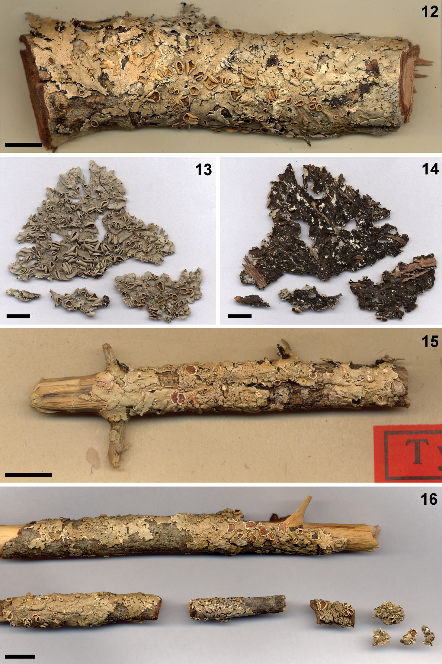
12 Lectotype of Parmelia amplectens 13 Holotype of Bulbothrix vainioi 14 Detail of the lower cortex of Bulbothrix vainioi 15 Holotype of Bulbothrix sensibilis 16 Holotype of Bulbothrix setschwanensis. Scale = 1 cm (14, 15, 16), 2 mm (17), 1 mm (18), and 20 µm (19).
Asia: India (
India, Mussoorie, northwest Himalaya, 7000 ft., leg. R. R. Stewart s.n., VII-1931 (NY 12298). Idem, Nilgherries Montains, Watt s.n. (lectotype of Parmelia amplectens, BM!, duplicate at GLAM!). Pakistan, Lower Topa, Murree hills, on bark of Pinus excelsa, leg. S. H. Iqbal 835, 11-VII-1967 (US). Malawi, Cholo Mt., dead branchlets of rain-forest trees, 1200 m alt., leg. L. J. Brass s.n. 24-IX-1946 (NY 17788). Brazil, São Paulo State, Itirapina Municipality, Estação Experimental de Itirapina, IF, leg. P. Jungbluth, L. S. Canêz & A. A. Spielmann PJ881, 26-III-2004. Idem, Santa Rita do Passa Quatro Municipality, Fazenda Vassununga, km 259 of the Anhanguera Highway, leg. M. P. Marcelli & B. L. Morretes 15653, 03-VI-1978 (SP). Idem, São Manuel Municipality, Fazenda Palmeira da Serra, non official particular cerrado (savannah) reserve, on tree trunk, leg. M. P. Marcelli & S. B. Barbosa 35693, 03-VI-2003 (SP). Idem, Botucatu Municipality, beside the highway that connects the city to the highway Castello Branco (SP-280), km 3, private cerradão forest inside Fazenda Morro do Ouro, 22°53'S, 48°26'W, 804 m alt, on a tree trunk, leg. M. P. Marcelli & S. B. Barbosa 35696, 4-VI-2003 (holotype of Bulbothrix vainioi, SP!).
The holotype (Fig. 11) consists of a single thallus on bark. It is partially detached from the substrate and in poor condition. Part of the upper cortex is absent, the medulla is much stained by oxidized salazinic acid, and the thallus is brittle and fragile. There are several apothecia in different stages of maturation, some of them also damaged, though they have ascospores. The thallus has many pycnidia, some containing conidia.
Nylander wrote on a label with the type specimen voucher “ascospores 14.0–18.0 × 7.0–11.0 mm”, but mentioned as measures 14.0–21.0 × 8.0–11.0 mm at the work in which he described Parmelia tiliacea var. meizospora (
Cilia in Bulbothrix meizospora are usually infrequent, and a portion of them in a same thalli apices might not present apices, while some others do not have bulbs. Often the bulbs become withered or reniform, which is more evident in the axillary cilia.
Regarding the presence and intensity of cortical maculae,
Apparently, as mentioned by
Bulbothrix setschwanensis (Zahlbruckner) Hale was compared to Bulbothrix meizospora by
Bulbothrix sensibilis (Steiner & Zahlbruckner) Hale was compared by
Bulbothrix hypocraea (Vainio) Hale was compared to Bulbothrix meizospora by
Recognition of Bulbothrix meizospora as a Bulbothrix species can sometimes be difficult, as commented already by
The type material (Fig. 12) of Parmelia amplectens Stirton (BM! lectotype, GLAM! duplicate) has cilia with more distinct bulbs and somewhat longer apices, and it is difficult to recognize maculae due its poor condition. However, the further characteristics agree with those of Bulbothrix meizospora as accepted by
Bulbothrix vainioi (Figs 13–14)Jungbluth, Marcelli and Elix was described by
Mycobank: MB 341612
Figure 15British East Africa, Bei-Bura (Kenia), auf Baumzweigen, leg. Schröder 285 (W!).
Thallus subirregularly to sublinearly sublaciniate, dusky gray in the herbarium, up to 6.9 cm diam., subcoriaceous, corticolous or ramulicolous; upper cortex 12.5−25.0 µm thick, algal layer 15.0−27.5 µm thick, medulla 87.5−120.0 µm thick, lower cortex 12.5−17.5 µm thick. Laciniae irregularly to occasionally anisotomically dichotomously branched, 1.3–5.2 mm wide, slightly imbricate, becoming crowded at the center, weakly adnate and loosely adpressed, with flat, subrounded to subtruncate apices; margins flat, slightly sinuous to crenate or irregular, entire to slightly incised, ocasionally sublacinulate; axils oval to irregular. Upper cortex smooth and continuous, becoming subrugose with occasional irregular cracks only on older parts; laminal ciliary bulbs absent. Adventitious marginal lacinulae scarce on older parts, short, 0.2–1.2 × 0.1–0.2 mm, plane, simple to irregularly branched; apices truncate; lower side concolor with the lower marginal zone. Maculae weak to distinct, puntiform, laminal, more evident at distal parts of the thallus. Cilia black, without or with simple and short apices, occasionally bent downwards, 0.05–0.20 (–0.30) × ca. 0.03 mm, with emerse bulbate bases 0.05−0.25 mm wide, occasionally withered and reniform, scarce along the margins, becoming frequent at the crenae and axils spaced ca. 0.05−0.15 mm from each other to eventually contiguous, solitary or in small groups becoming absent or scarce at the apices and adjacent parts of the laciniae. Soredia, Isidia, and Pustulae absent. Medulla white. Lower cortex black, with random dark brown spots at the transition to the center, slightly shiny, smooth to subrugose or subvenate, moderately rhizinate. Marginal zone mostly brown, attenuate, ca. 0.5−2.0 mmwide, partially black and indistinct from the center, slightly shiny, smooth to subvenate, weakly rhizinate until the transition to the center. Rhizinae black, sometimes partially dark brown close to the margins, simple to rarely furcate, without basal or displaced bulbs, 0.10–0.30 (-0.40) × ca. 0.05 mm, usually frequent but scarcer at the margins and at the transition to the center, evenly distributed. Apothecia concave to subplane, sessile to adnate, 0.2−4.3 mm diam., laminal, ecoronate; margin and amphitecia initially smooth becoming subrugose, without ornamentations. Disc pale brown, epruinose, imperforate; epithecium 10.0–17.5 µm high; hymenium 30.0−47.5 µm high; subhymenium 20.0−30.0 µm high. Ascospores ellipsoid to oval, (7.0−) 8.0−12.0 (−13.0) × 5.0−7.0 µm; epispore ca. 0.75 µm. Pycnidia frequent, laminal, immersed, with black ostioles. Conidia baciliform to weakly bifusiform 5.0−9.0 × 0.75 µm.
TLC/HPLC: cortical atranorin, medullary salazinic and consalazinic acids (see also Hale 1976).
Asia: Sri Lanka (
Venezuela, Táchira, Via Rubio, Brámon, 800–1100 m, leg. M. E. Hale & M. López Figueiras 45727, 24-III-1975 (US). Brazil, São Paulo State, 6 km SW of Jaboticabal, 21°35'S, 48°35'W, on trees in cerradão, leg. A. Fletcher 10138, 03-V-1975 (BM). Idem, Pirassununga, Rawitscher Reserve, Cerrado auf Zweigen, leg. H. Walter & E. Walter Br 58, 30-IX-1965 (M).
The holotype of Bulbothrix sensibilis (Fig. 15) consists of a small thallus ca. 6.0 cm in diameter on tree branch, in a reasonable state of preservation, although several parts and apothecia are badly damaged. The material is glued to the card voucher, and it was necessary to free some laciniae for observation of the lower cortex. There are apothecia containing ascospores in good condition and there are several pycnidia with conidia.
Steiner and Zahlbruckner (
The material atributted by
It is possible that
Bulbothrix hypocraea (Vain.) Halediffers by being more evidently maculate than Bulbothrix sensibilis, by the pale brown lower cortex with slighly darker margins, and by the brown rhizines with dark basal or displaced bulbs.
Bulbothrix bulbochaeta (Hale) Hale (LWG! holotype, US! isotype) differs by the narrower laciniae ca. 1.0−2.5 mm wide, the branched cilia and rhizines, the constant presence of laminal ciliary bulbs, the coronate apothecia containing very small and rounded ascospores 4.0−6.0 × 3.0−4.0 µm and by the absence of medullary substances.
Bulbothrix linteolocarpa Marcelli was compared to Bulbothrix sensibilis by
Bulbothrix meizospora (Steiner & Zahlbruckner) Hale differs by the laciniae usually more irregularly branched and with rounded apices, and by the always larger ascospores, measuring 12.0−22.0 × 8.0−12.0 µm. Comparatively, thalli of Bulbothrix sensibilis are also more evidently maculate.
Mycobank: MB 341613
Figure 16China, Prov. Setschwan austro-occid., in regionis siccae subtropicae convallis fluminis Yalung ad septentriones oppidi Yneyünen infra castelum Kwapi ram Pistacia weinmannifolia supra vic. Otang, alt. 2400–2500 m., leg. Handel-Mazzetti 2739, 30-V-1914 (WU!).
Thallus subirregularly to sublinearly laciniate, greenish gray in the herbarium, up to 7.0 cm diam., subcoriaceous, corticolous or ramulicolous; upper cortex 15.0−20.0 µm thick, algal layer 30.0−47.5 µm thick, medulla 87.5−110.0 µm thick, lower cortex 12.5−20.0 µm thick. Laciniae irregularly to partially to anisotomically dichotomously branched, contiguous to imbricate, 1.1–3.5 (-5.0) mm wide, adnate and adpressed, with ±flat, subrounded to subtruncate apices; margins flat, smooth and sinuous to crenate or or irregular, entire to slightly incised, occasionally sublacinulate; axils oval or irregular. Upper cortex mostly smooth and continuous, occasionally becoming subrugose and irregularly cracked; laminal ciliary bulbs absent. Adventitious marginal lacinulae scarce on older parts, short, 0.2–1.0 × 0.1–0.6 mm, plane, simple to irregularly branched; apices truncate or acute; lower side concolor with the lower marginal zone. Maculae absent. Cilia black, without or with simple apices, 0.05–0.30 (–0.50) × ca. 0.03 mm, with semi-immersed to emerse bulbate bases 0.05–0.25 mm wide, frequent to abundant along the margins, spaced 0.05−0.15 mm from each other to rarely contiguous, solitary or in small groups at the crenae and axils becoming scarce at the apices of the laciniae. Soredia, Isidia and Pustulae absent. Medulla white. Lower surface pale brown, opaque, smooth to subrugose, moderately rhizinate. Marginal zone pale brown, indistinctly delimited from the center, opaque, smooth to subrugose, weakly papillate, variably rhizinate. Rhizines brown or cream colored, simple, rarely with subtle displaced blackish bulbs, 0.05–0.80 × 0.03–0.05 mm, frequent becoming abundant near the margins, evenly distributed. Apothecia subconcave to plane, adnate to subpedicelate, 0.4−4.1 mm diam., laminal to submarginal, ecoronate; margin smooth to subcrenate or fissured; amphitecia smooth, without ornamentations. Disc light to dark brown, epruinose, imperforate; epithecium 7.5–12.5 mm high; hymenium 35.0−42.5 µm high; subhymenium 12.5−20.0 µm high. Ascospores ellipsoid to oval, (10.0−) 12.0−19.0 × 6.0−9.0 (−10.0) µm; epispore ca. 1.0 µm. Pycnidia laminal, frequent, immerse, with black ostioles. Conidia baciliform to weakly bifusiform (4.0−) 5.0−8.5 × ca. 0.75 µm.
TLC/HPLC: cortical atranorin and chloroatranorin, medullary salazinic and consalazinic acids (see also Hale 1976).
Asia: China (
India, Oriental India, prov. Central, Chavradadar, Manra distr., 3500 ft., leg. J.Masten s.n., XII-1900 (NY). Idem, Kolhapur, Maharashtra, Panhala Forest, leg. P. G. Pahvardhan & R. A. V. Prabhu 74.1007, 13-X-1974 (US). Idem, Índia, E. Himalayas, Darjeeling, West Bengal, Manibhanjan, 7700 ft., leg. C. G. Dharne & K. N. R. Chaudhuri 82, VI-1966 (SP). Pakistan, Lower Topa, Murree hills, on bark of Pinus excelsa, leg. S. H. Iqbal 844(?), 11-VII-1967 (US).
The holotype (Fig. 16) consists of a thallus on a tree twig, together with other bark fragments containing smaller pieces. It is in a reasonable state of preservation, with some lobes and apothecia badly damaged. The material contains several apothecia at different stages of maturity with ascospores in good condition, and many pycnidia with conidia. There are some loose fragments, on which the lower cortex was observed.
Among similar species, Bulbothrix meizospora is morphologically close to Bulbothrix setschwanensis including the ascospores of similar size, but the has a distinct black lower cortex with brown margins, as cited by
Bulbothrix continua (Lynge) Hale differs by the narrower laciniae ca. 1.0−2.0 mm wide and by the smaller ascospores 9.0−13.5 × 5.0−7.5 µm. in direct comparison, morphologically its aspect more closely resembles that of Bulbothrix hypocraea, although the maculations are absent, while that of Bulbothrix setschwanensis is more akin to that of Bulbothrix meizospora. In a key in
Originally described from China, the species is also known from India and Nepal (
The author wishes to thank the curators of BM (Scott LaGrecca), FH (Donald Pfister), GLAM (Keith Watson), H (Leena Myllys), HUFSCAr, LD (Arne Thell), LG (Emmanuël Sérusiaux), M (Andreas Beck), NY(Barbara Thiers), S (Anders Tehler), TNS (Yoshihito Ohmura), TUR (Seppo Huhtinen), US (Rusty Russell), W (Uwe Passauer) and WU (Walter Till) for the loan or disposition of the type specimens and additional material, Dr. John A. Elix for HPLC data on the species substances, Dr. Harrie Sipman for the English review, comments, and suggestions, and the reviewers for critical revision of the manuscript. Open access to this paper was supported by the Encyclopedia of Life (EOL) Open Access Support Project (EOASP).