|
||
|
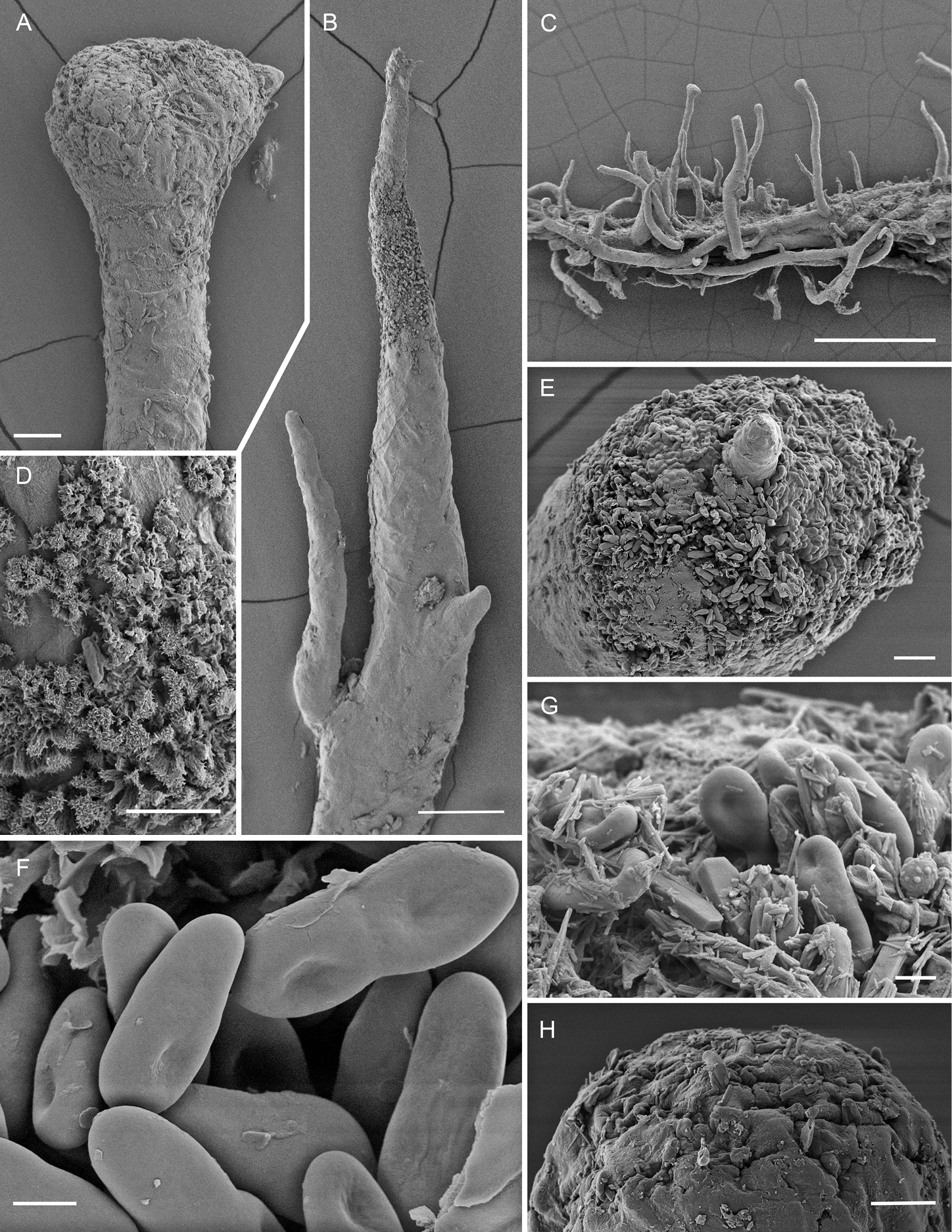
Scanning electron micrographs of Chaenothecopsis matai sp. nov. (PDD 110749) A semi-mature capitulum B upper part of apothecium C pseudostroma-like growth of apothecia D structure of pruina on stipe surface E proliferating growth of capitulum F ascospores G (detail of E): ascospores and crystals on capitulum surface H mature capitulum. Scale bars: 1 mm (C); 100 µm (B); 30 µm (A); 20 µm (E); 10 µm (D, H); 2 µm (F, G). |