|
||
|
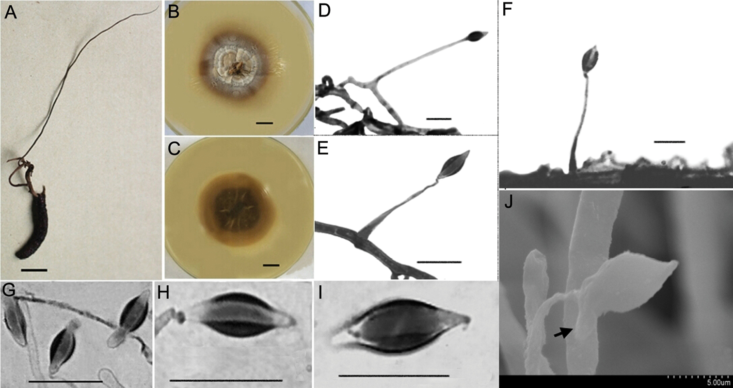
Morphological characteristics of Hirsutella kuankuoshuiensis A the insect specimens with single and thin synnemata (HKAS112885) B, C colonial morphology on PDA agar media for 20 d B shows the front of the colony and C shows the back of the colony D–G LM images showing conidiogenous cells and conidia D, E the structure of conidiogenous cells on mycelia F the images of conidiogenous cells on synnemata (optical microscope) H–J conidial morphology (LM) G conidia with mucilage (SEM). Scale bars: 10 mm (A–C); bar of G was shown in the figure; the rest of the bars were 10 μm. LM, light microscopy; PDA, potato dextrose agar; SEM, scanning electron microscopy. |