|
||
|
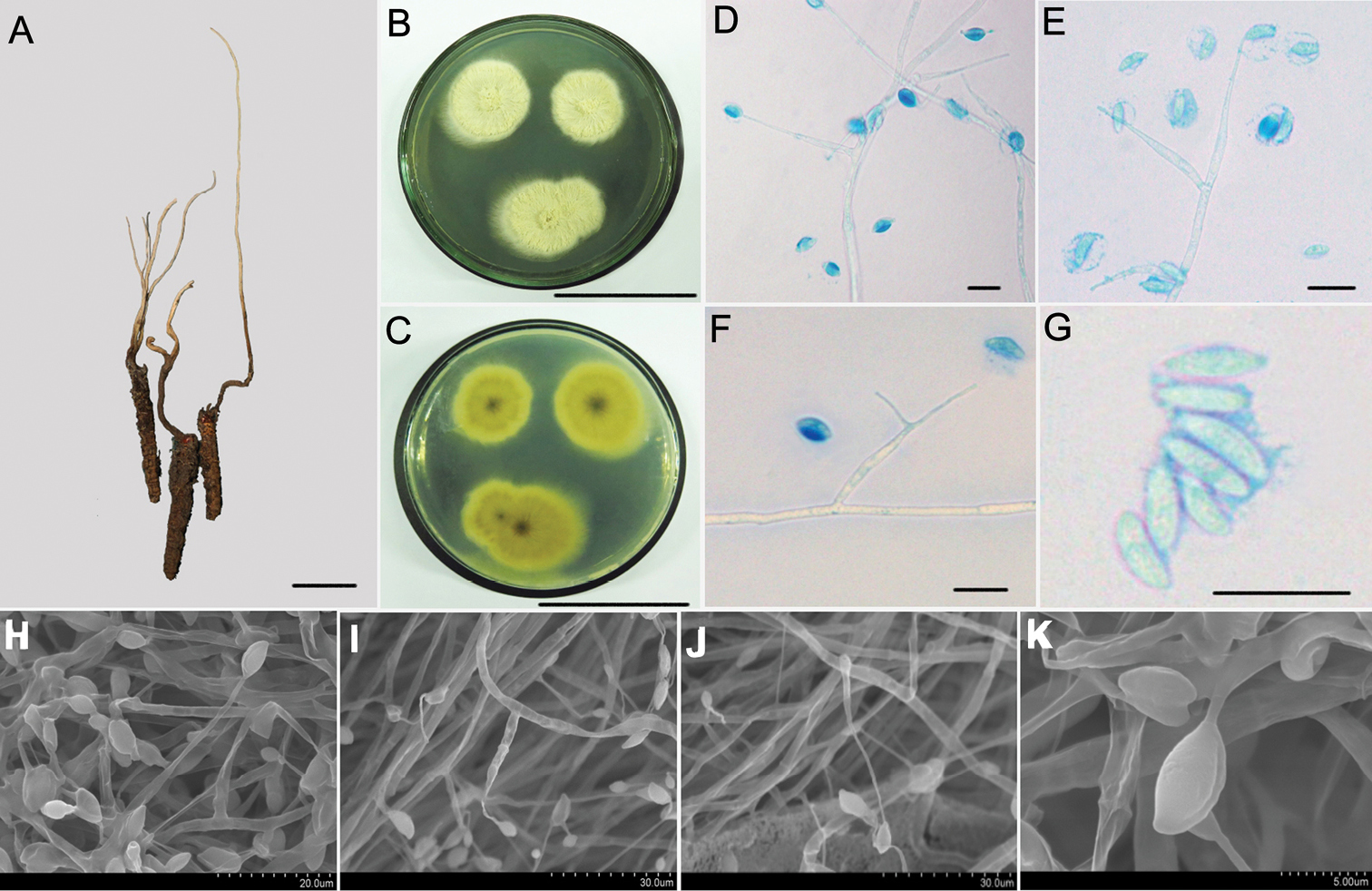
Morphological characteristics of Hirsutella flava A the infected insect specimens with a long and single synnemata (HKAS112884) B, C colonial morphology on PDA agar media for 20 d B shows the front of the colony and C shows the back of the colony D–G LM images of the general morphology of conidiogenous cells and conidia H–K SEM images showing conidiogenous cells and conidial structure; Scale bars: 1 cm (A); 5 cm (B, C), 10 μm (D–G); the rest of the bars are shown in the figure. LM, light microscopy; PDA, potato dextrose agar; SEM, scanning electron microscopy. |