|
||
|
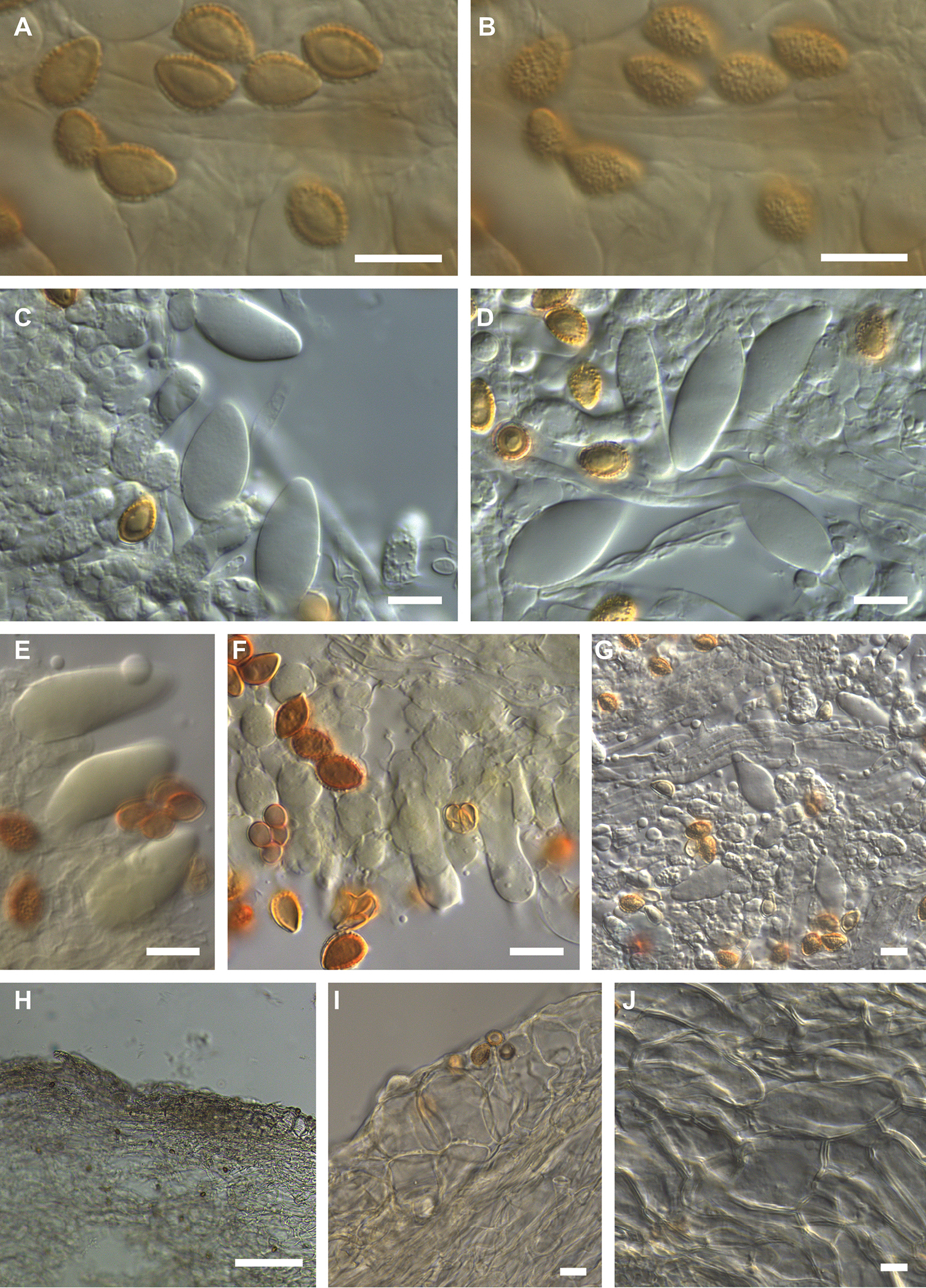
Microscopic features of Hebeloma radicans holotype (E. Horak 13265) A spores in KOH ×1600 B spore ornamentation in KOH ×1600 C cheilocystidia and basidium in KOH ×1000 D cheilocystidia and basidium in KOH ×1000 E pleurocystidia in Melzer’s reagent ×1000 F basidia in KOH ×1000 G pleurocystidia in KOH ×500 H sectional view of ixocutis showing thin gelatinous epicutis in KOH ×125 I sectional view of subcutis and trama below subcutis in KOH ×500 J sectional view of trama below subcutis in KOH ×500. Scale bars: 10 µm, 100 µm (H). Photographs H.J. Beker. |