|
||
|
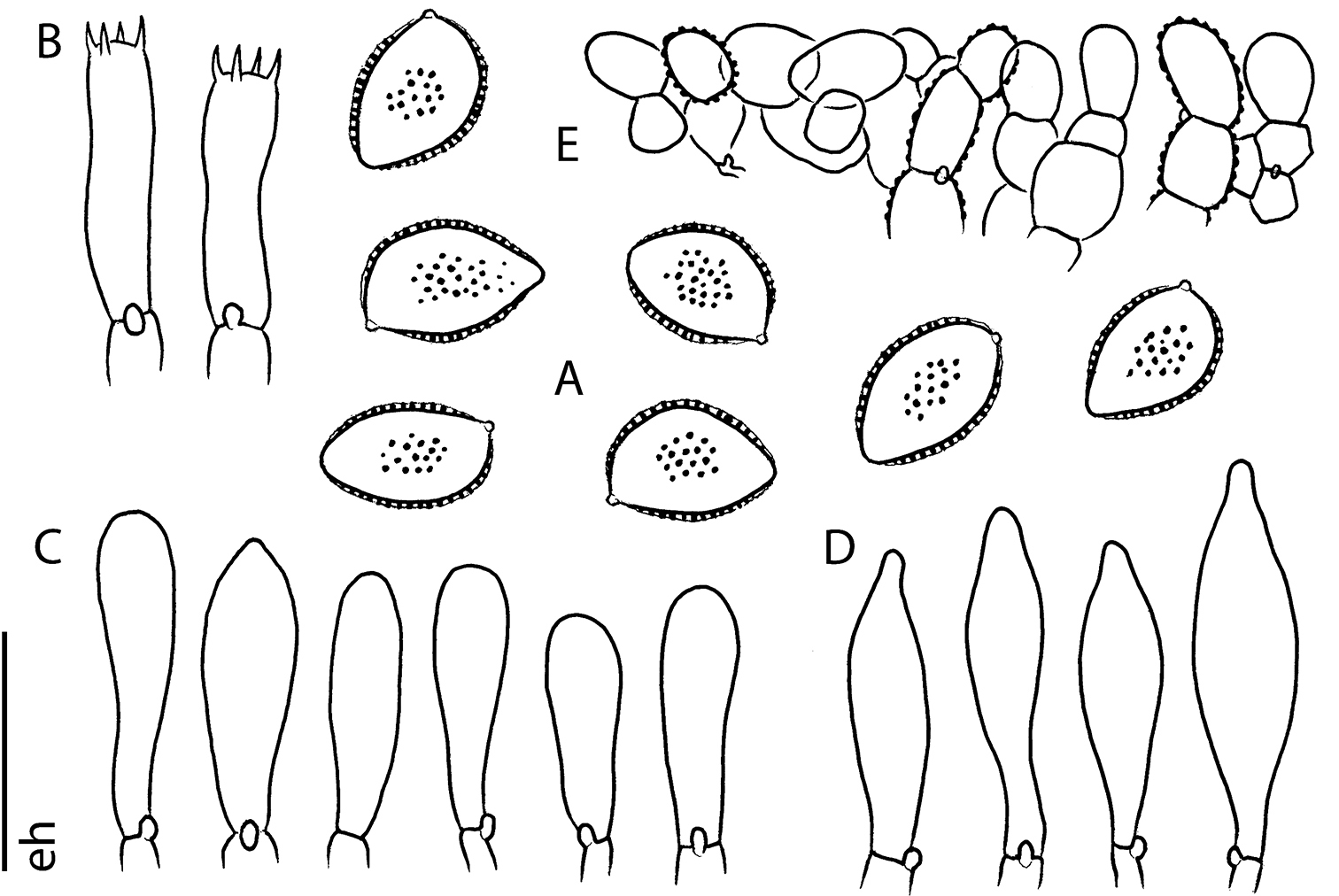
Microscopic features of Hebeloma flavidifolium (E. Horak 13406) A spores ×2000 B basidia ×1000 C cheilocystidia ×1000 D pleurocystidia ×1000 E pileipellis (section of subcutis below epicutis) ×500. Scale bar: 10 µm ×2000, 20 µm ×1000 and 40 µm ×500. Drawing E. Horak. |