|
||
|
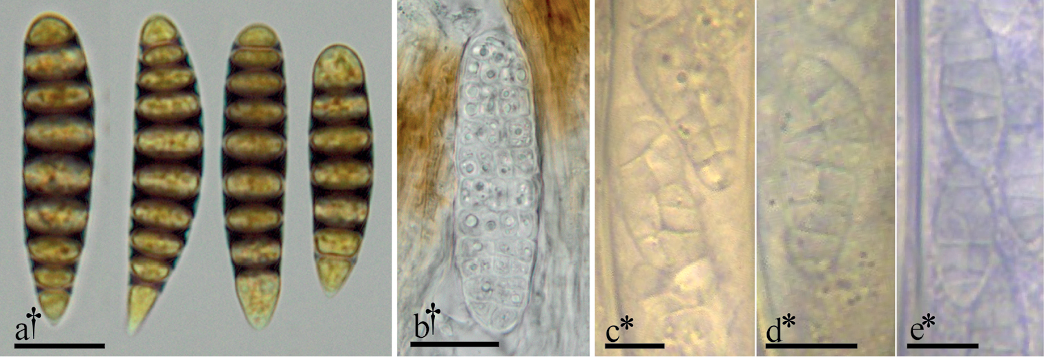
Select examples of ascospore morphologies in Graphidaceae and Leotiomycetes a ascospores of Glyphis cicatricosa in Lugol’s solution b muriform ascospore of Mellitiosporium versicolor c–e muriform ascospores of Claussenomyces spp. within living, immature asci. All microphotographs of cells and tissues mounted in water unless otherwise noted. † = dead, * = living. Scale bars: 10 µm (a); 20 µm (b); 5 µm = (c–e). Specimens photographed: a = J.M.K personal collection; b = U.S.A., Oregon, Horse Rock Ridge, M. A. Sherwood, L. H. Pike & D. Wagner, 21 Mar 1979, FH [s.n.], image courtesy of Farlow Herbarium of Harvard University; c–e = L.Q. personal collections. |