|
||
|
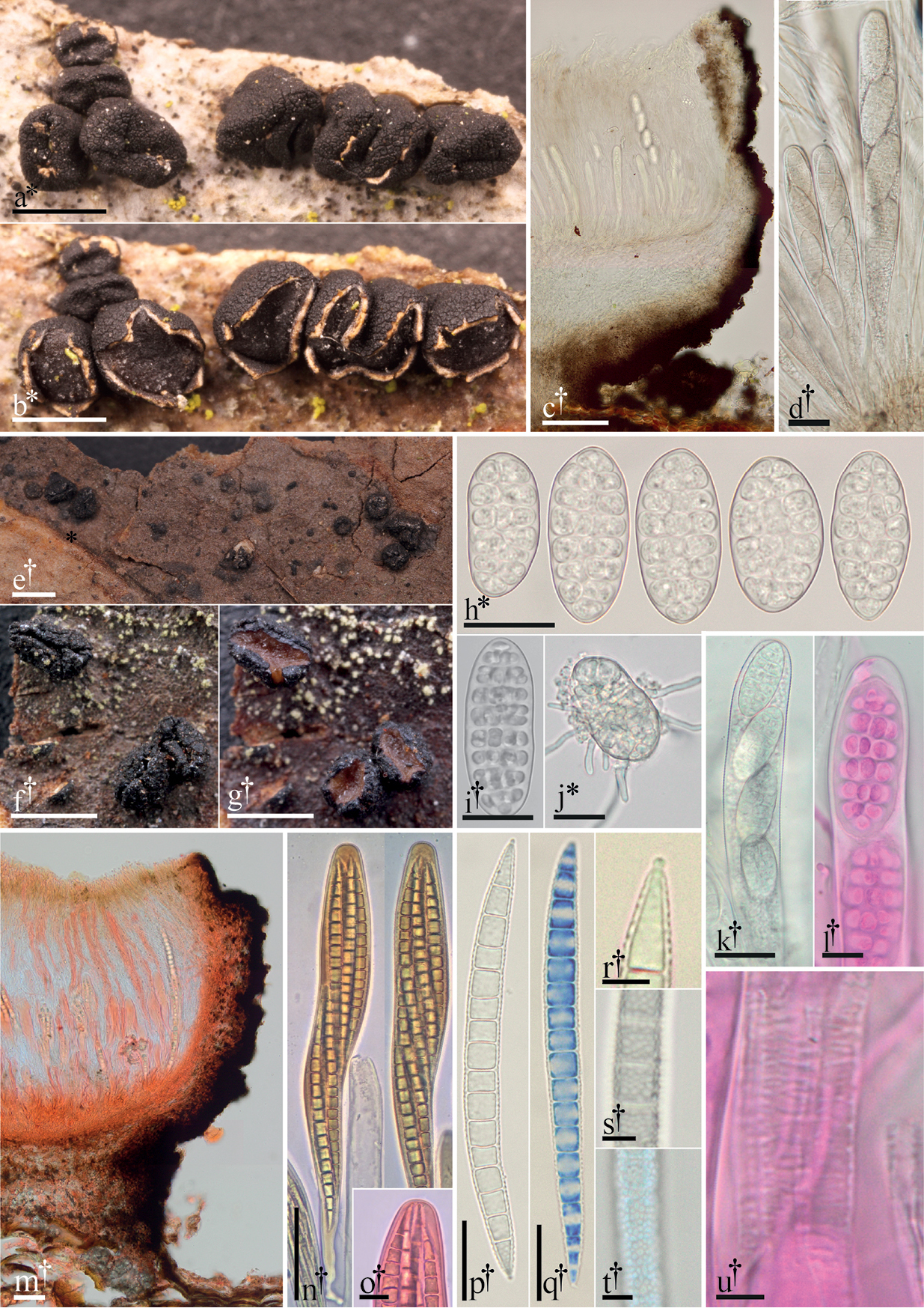
Morphological features of Triblidiaceae. a–d, h–l, u Triblidium caliciiforme a dried apothecia on bark b same apothecia hydrated c 15 µm thick longitudinal section d dead asci containing living ascospores h–i ascospores j germinating ascospore k dead ascus containing living ascospores, detailing the apex l ascus detailing the apex (phl) u fine transverse striations of dehisced ascus (phl). e–g, m–t Huangshania verrucosa e habit of apothecia on bark (dried) f detail of dried apothecia g detail of same apothecia hydrated m 15 µm thick longitudinal section in (Cr) n asci (KOH & Mlz) o detail of ascus apex (Cr) p ascospore q ascospore (cb/l) r detail of plug-like structure in a terminal cell of an ascospore s–t detail of verrucose ascospore surface (t in cb/l). All microphotographs of cells and tissues mounted in water unless otherwise noted: Congo red (Cr), cotton blue in lactophenol (cb/l), Melzer’s reagent (Mlz), phloxine (phl), potassium hydroxide (KOH). † = dead, * = living. Scale bars: 1 mm (a–b, e–g); 50 µm (c, m–n); 20 µm (h–k, p–q); 10 µm (l, o); 5 µm (r–u). Specimens photographed: T. caliciiforme: a–b, d, j GJO-0088904; i FH-15071105; c, h, k–l, u CUP-18080101; H. verrucosa: UME-29336a. |