|
||
|
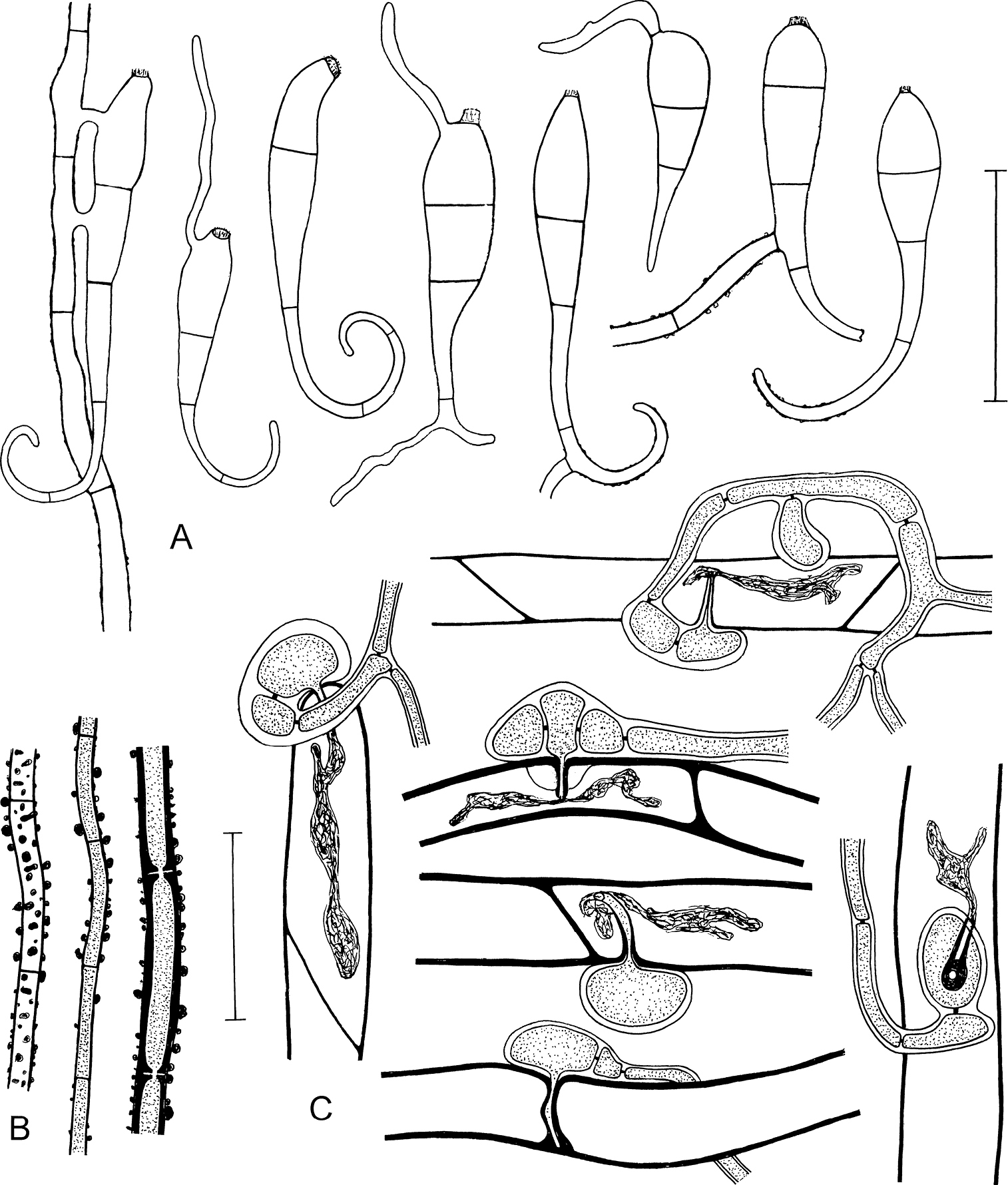
Microscopic characters of Octospora conidiophora agg. (lineage B). A Conidia, distal curved part apparently sometimes broken off, five conidia germinating by formation of usually a single hypha, conidium on the left connected to a hypha by two anastomoses B Strongly warted hyphae, the left one seen from above, the two others in optical section C Appresssoria infecting rhizoids in lateral view, the right one seen from above, infection pegs surrounded by lignituber-like tubes formed by the host cell wall, intracellular haustoria present apart from the lowermost infection where the peg is completely encapsulated by the host cell wall A, B, C ZE37/18. Scale bars: 50 µm (A); 30 µm (B, C). Illustrated by P.D. |