|
||
|
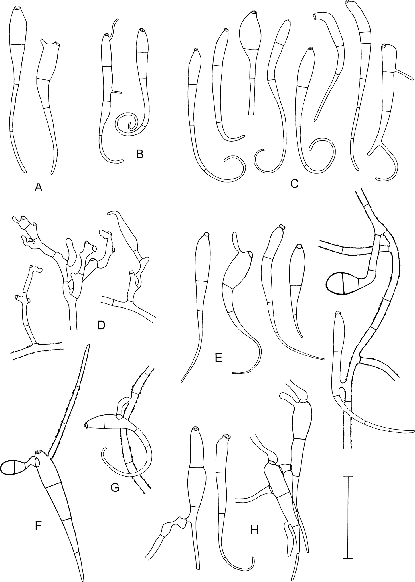
Microscopic characters of Octospora conidiophora. A–C, E–H Conidia, distal curved part apparently sometimes broken off, some conidia germinating D Conidogenous cells, on the right with a developing conidium E (on the right) Conidium anastomosing to mycelial hypha with two-celled appressorium F Conidium germinating by a hypha with a warty surface and a two-celled appressorium G Conidium with anastomosis to mycelial hypha H (on the right) Two germinating conidia with an anastomosis between them A, F ZE63/18 B ZE46/18 C ZE77/18 D, E holotype ZE48/18 G ZE57/18 H ZE11/18. Scale bar: 50 µm. Illustrated by P.D. |