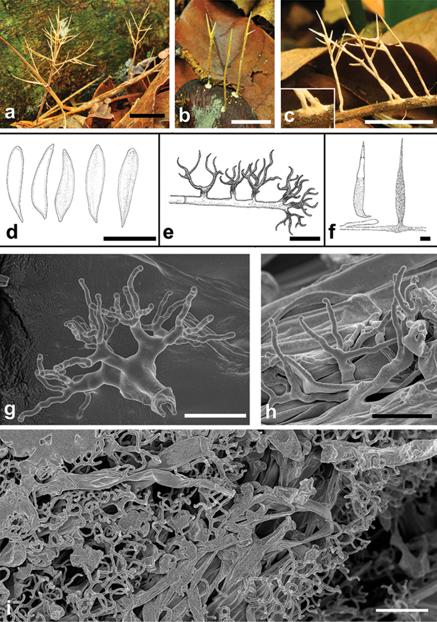
Parapterulicium subarbusculum: a–c basidiomes in the field. The detail in c shows the developing corticioid patch d basidiospores e dichophyses f gloeocystidia g, hSEM images of dichophyses; i. SEM images of basidiome surface with abundant dichophyses. Scale bars: a–c = 1 cm; d–f, i = 10 μm; g, h = 5 μm.


  Part of: Leal-Dutra CA, Neves MA, Griffith GW, Reck MA, Clasen LA, Dentinger BTM (2018) Reclassification of Parapterulicium Corner (Pterulaceae, Agaricales), contributions to Lachnocladiaceae and Peniophoraceae (Russulales) and introduction of Baltazaria gen. nov. MycoKeys 37: 39-56. https://doi.org/10.3897/mycokeys.37.26303