|
||
|
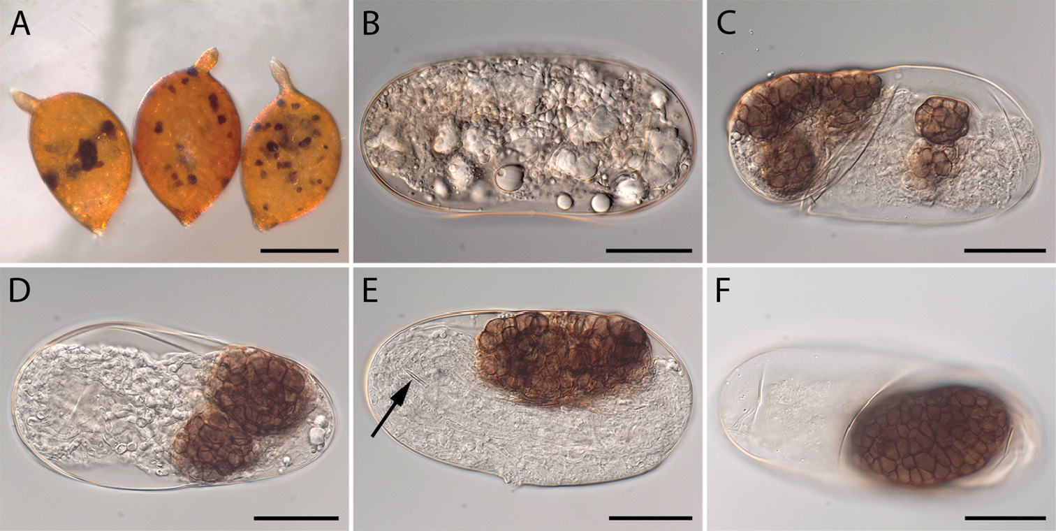
Cysts and eggs of Heterodera filipjevi infected by Monocillium bulbillosum in vitro. A Symptomatic cysts infected by M. bulbillosum B Infected egg showing early stage of fungal colonisation C–F Formation of microsclerotia in nematode eggs (arrow points at nematode’s stylet). Scale bars: 300 µm (A); 30 µm (B–F). |