|
||
|
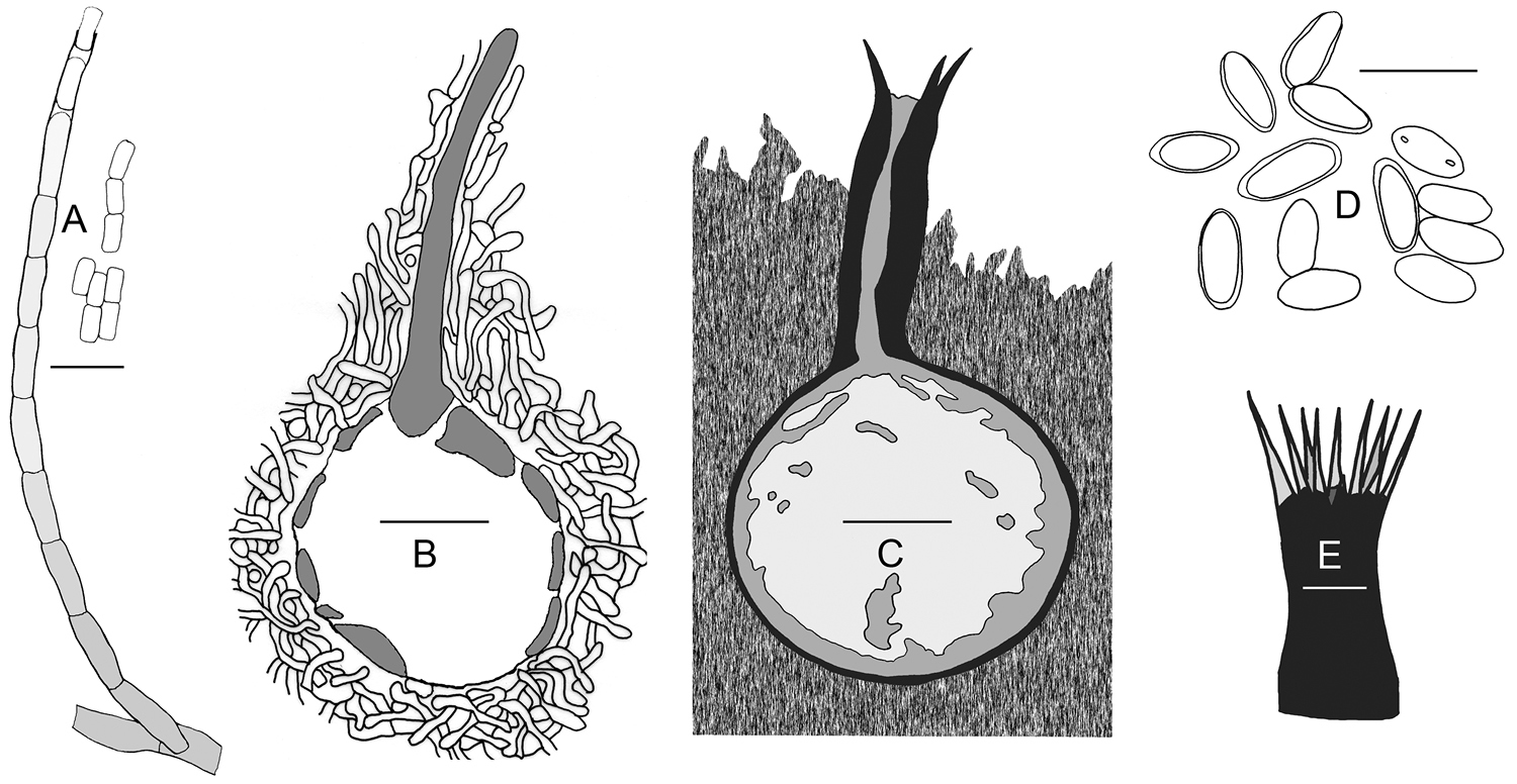
Line drawings of the oak wilt fungus. These illustrations are based on previously published line drawings and observations of the herbarium specimens (BPI 595712, FP 97476) in the present study. A Conidiophore and conidia in 10 % KOH (BPI 595712) B Ascomatal primordium re-drawn from Wilson (1956)C Median, histological section through ascoma embedded in the mycelial mat, re-drawn from Bretz (1952)D Ascospores in 10 % KOH (FP 97476) E Ostiolar hyphae (FP 97476). Scale bars: A, D = 10 µm, E = 50 µm, B, C = 100 µm. |