|
||
|
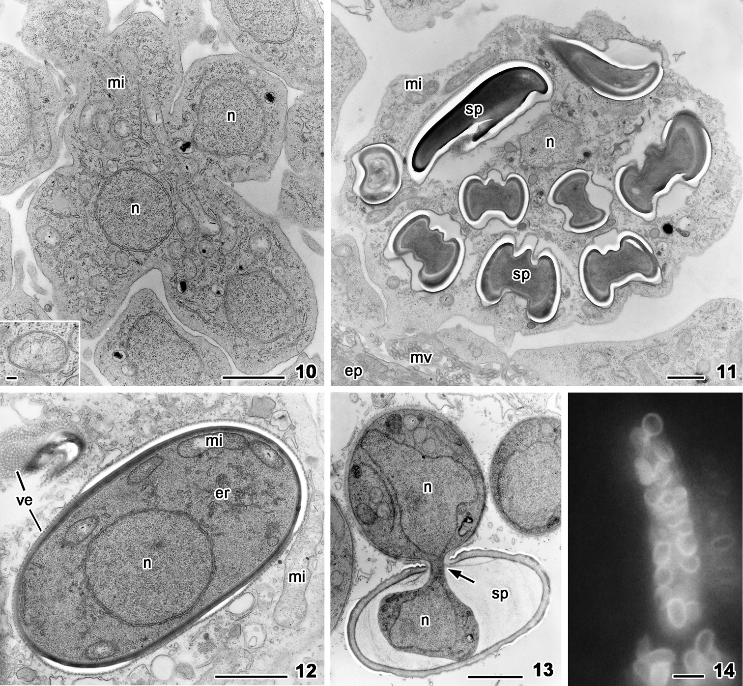
Nephridiophaga blattellae, 10–13 transmission electron microscopy, 14 Calcofluor white staining. 10 Meront with several nuclei (n) and mitochondria (mi) in the lumen of Malpighian tubule. Inset: Mitochondrium with tubular to sac-like cristae. 11 Sporogenic plasmodium containing mature spores (sp), mitochondria (mi), and vegetative nuclei (n) in the cytoplasm. The plasmodium is anchored to the microvilli (mv) of epithelial cells (ep) of the tubule. 12 Young spore within the cytoplasm of a sporogenic plasmodium, surrounded by a layer of vesicles. The spore cytoplasm contains one nucleus (n), mitochondria (mi), and endoplasmic reticulum (er). 13 An infectious sporoplasm hatches through the central spore opening, leaving behind the spore wall of the emptying spore (sp). The nucleus (n) is squeezed through the tiny spore opening. 14 Calcofluor white stains the spore wall indicating the presence of chitin (bluish color). Scale bars: 1 µm (10–13), inset 0.1 µm (10), 5 µm (14). |