|
||
|
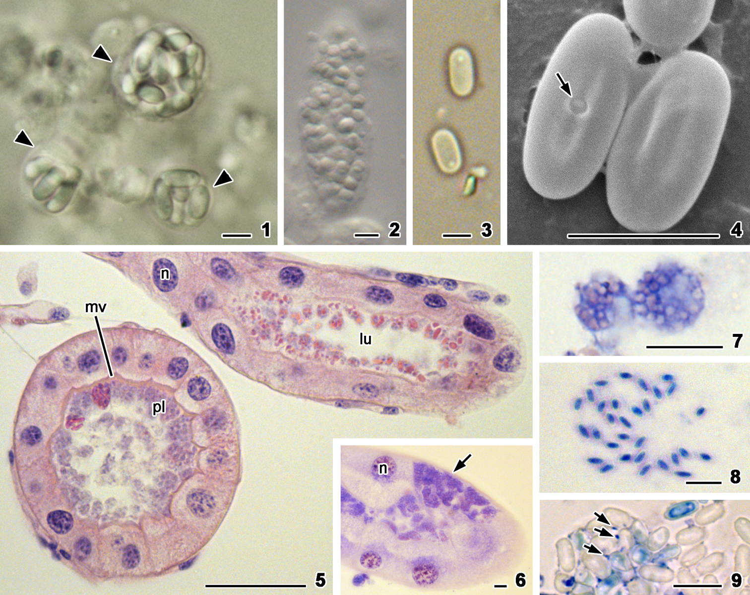
Nephridiophaga maderae, 1, 2, 5–9 bright field 3 phase contrast 4 scanning electron microscopy. 1 Three sporogonial plasmodia with different numbers of included spores. Arrows point to plasma membrane. 2 Merogonial plasmodium with numerous nuclei. 3 Mature spores. 4 The upper surface of the spore possesses a central spore opening (arrow, left spore) while the lower surface of the spore lacks an opening (right spore). 5, 6 Paraffin sections stained with hematoxylin-eosin. Generally, the plasmodia (pl) are found in the lumen of the Malpighian tubule but are often attached to the microvilli (mv) (5). Rarely, aggregates of vegetative plasmodia (arrow) occur in the epithelial cells of the Malpighian tubules (6). n = nuclei of epithelial cells. 7–9 Smears of macerated tubules stained with Giemsa depicting vegetative plasmodia (7), stained young spores (8), and unstained mature spores with residual nuclei (arrows) of the mother sporoplasm. Scale bars: 5 µm (1–4), 50 µm (5), 10 µm (6–9). |