|
||
|
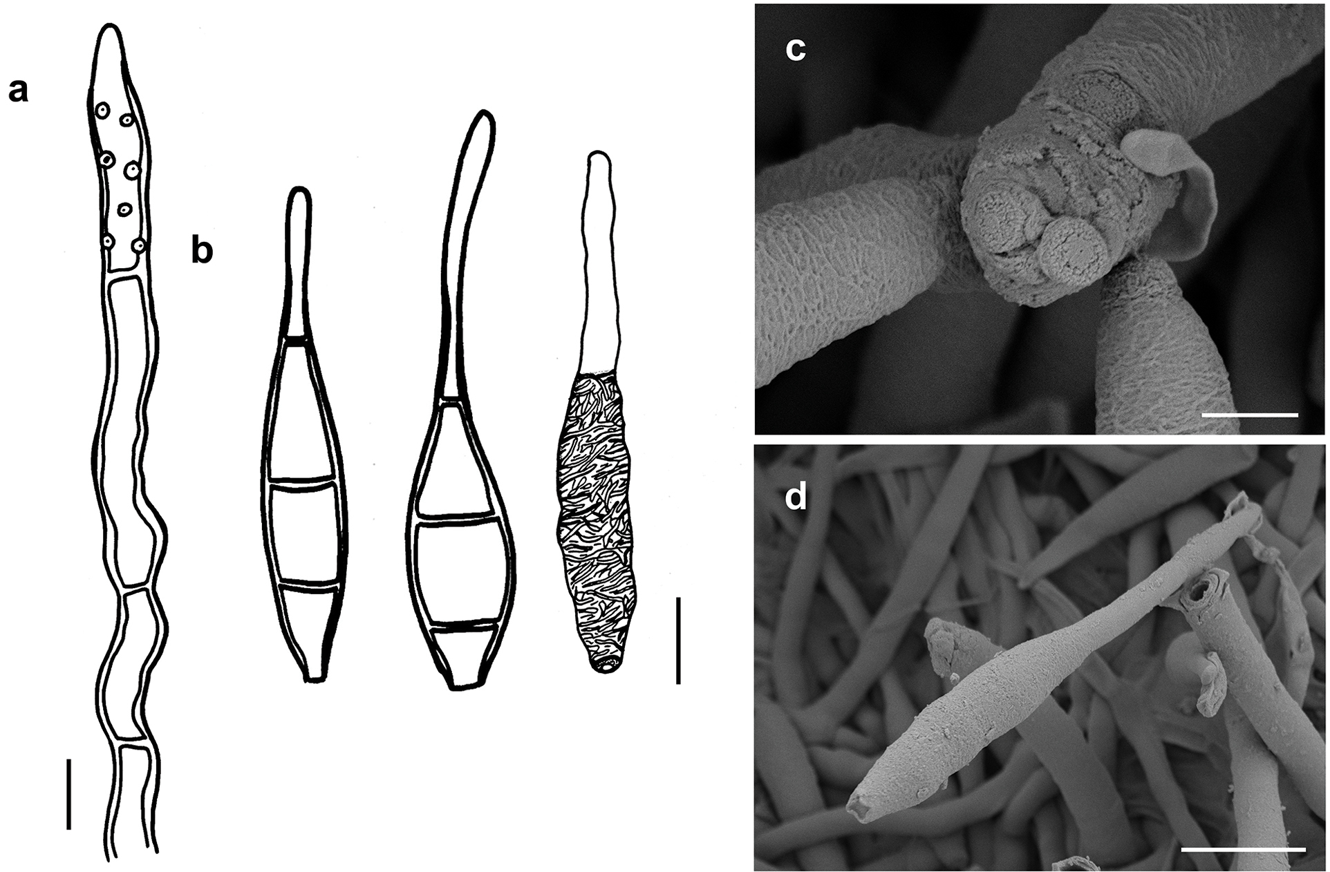
Spiropes effusus (IMI 130721) a conidiophore shown in optical section b conidia. The first two drawings show spores in optical section. The right-hand drawing shows a conidium as seen by SEM c, d as seen by SEM c conidiophore with scars and conidia d conidium. Scale bars: 5 μm (a); 8 μm (b); 2 μm (c); 8 μm (d). |