|
||
|
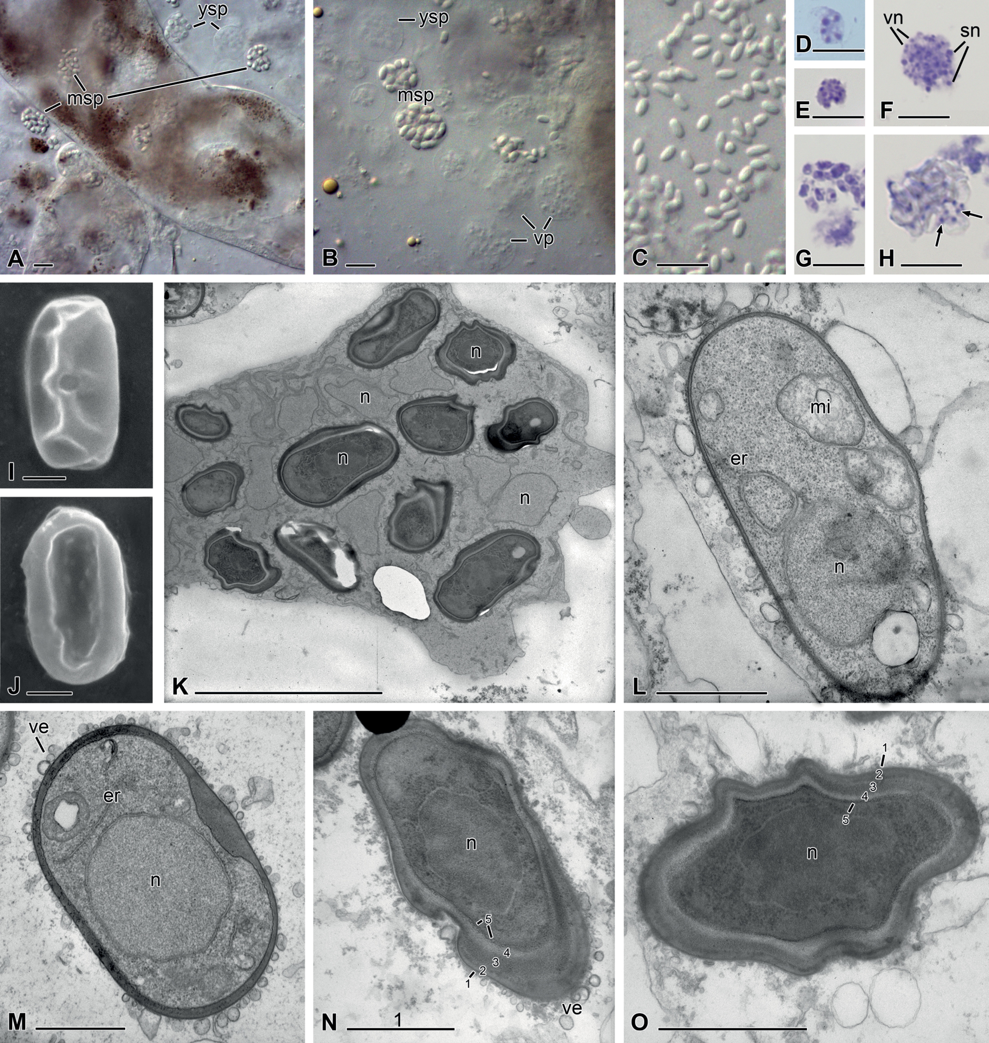
Nephridiophagid (Malpighivinco podagricae) from Podagrica malvae A–C differential interference microscopy (DIC) D–H Giemsa staining I, J scanning electron microscopy (SEM) K–O transmission electron microscopy (TEM) A fresh squash preparation of infected Malpighian tubules with young sporogenic plasmodia (ysp) and mature sporogenic plasmodia (msp) inside and outside the tubules B nuclei of vegetative plasmodia (vp) and young spores in sporogenic plasmodia have less contrast than mature spores C mature spores D, E vegetative plasmodia with few (D) or many (E) nuclei F young sporogenic plasmodium with small vegetative nuclei (vn) and larger sporogenic nuclei (sn) G the nuclei of thin-walled young spores are stained H in mature sporogenic plasmodia, only the vegetative nuclei between the spores are stained (arrows) I, J flattened mature spores with central spore opening on one side (I) K ultra-thin section of a sporogenic plasmodium with uninucleated mature spores and vegetative nuclei in the mother cytoplasm. n = nuclei L thin-walled young spore with a nucleus in polar position, mitochondria (mi) and endoplasmic reticulum (er) M maturing spore with attached vesicles (ve) delivering spore wall material and centrally located nucleus N, O mature spores with five-layered, thick spore wall, dense cytoplasm and central nucleus. Scale bars: 10 µm (A–H); 5 µm (K); 1 µm (I, J, L–O). |