|
||
|
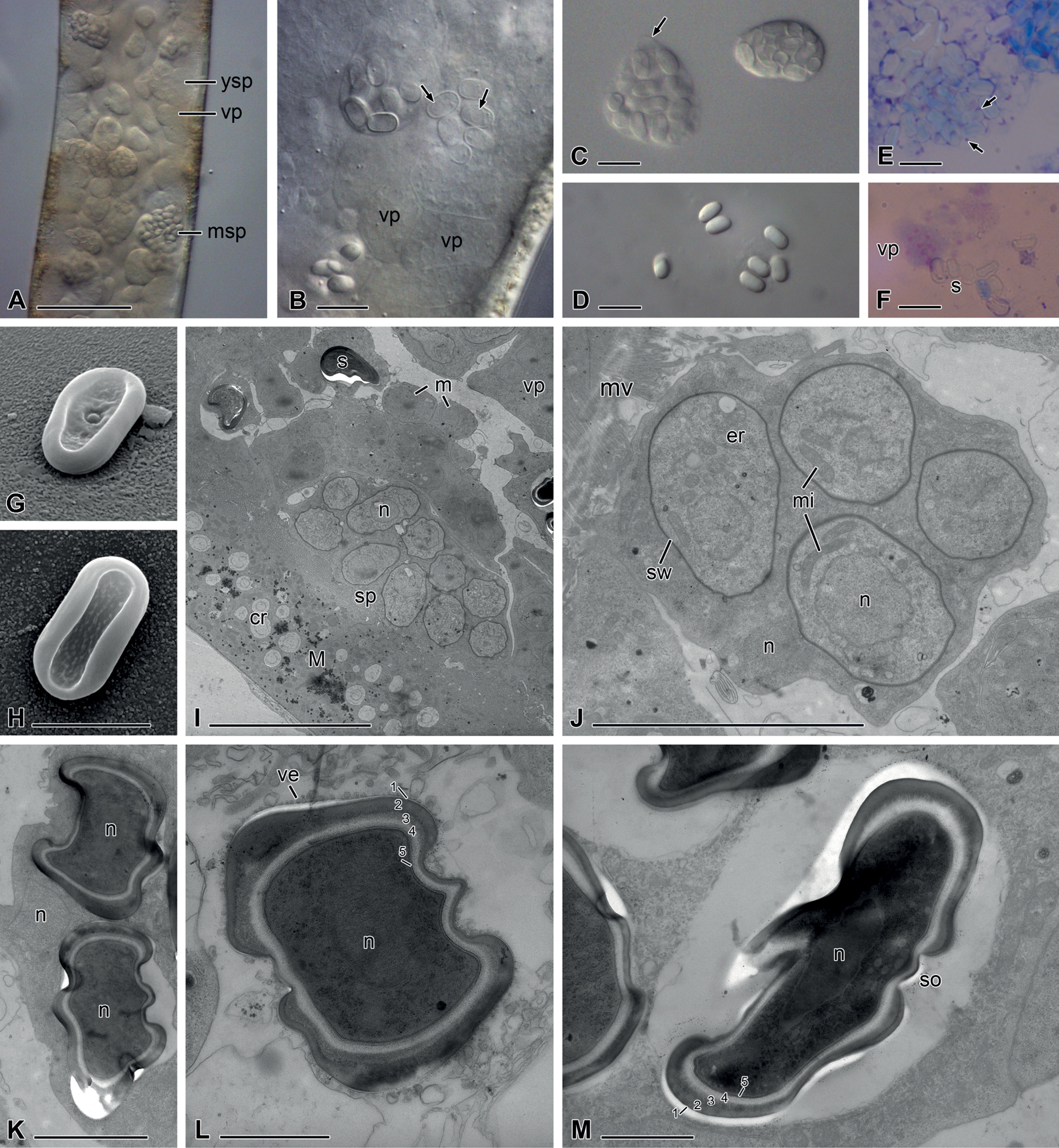
Nephridiophagid (Nephridiochytrium forficulae) from Forficula auricularia A–D differential interference microscopy (DIC) E, F Giemsa staining G, H scanning electron microscopy (SEM) I–M transmission electron microscopy (TEM) A Malpighian tubule filled with vegetative plasmodia (vp), young sporogenic plasmodia (ysp) and mature sporogenic plasmodia (msp) B young spores have a thin, yet transparent spore wall and a nucleus positioned near a cell pole or centrally C left plasmodium with large mature spores, right plasmodium with smaller, younger spores. Arrow points to plasma membrane D single mature spores E Giemsa staining reveals residual nuclei (arrows) of the plasmodium between mature spores F vegetative plasmodium with stained nuclei. s = spores G, H flattened oval spores with rim and a central spore opening on one side (G) I ultrathin section through a Malpighian tubule infected with different stages: small, uninucleate merozoites (m), vegetative plasmodia with several nuclei, sporogenic plasmodia (sp) and mature spores. n = nucleus. The epithelium of the Malpighian tubule (M) contains concretions (cr) J young sporogenic plasmodium containing young spores with a thin spore-wall (sw), one nucleus, endoplasmic reticulum (er) and mitochondria (mi) K part of a mature sporogenic plasmodium with a residual nucleus and two mature spores with a centrally located nucleus and a thick spore-wall L cross-section of a mature spore in the degenerating plasmodial cytoplasm. The spore wall consists of five layers (1–5). ve = vesicles M mature spore in longitudinal section showing the thin-walled cap of the spore opening (so). Scale bars: 50 µm (A); 10 µm (B–F, I); 5 µm (G, H, J); 2 µm (K); 1 µm (L, M). |