|
||
|
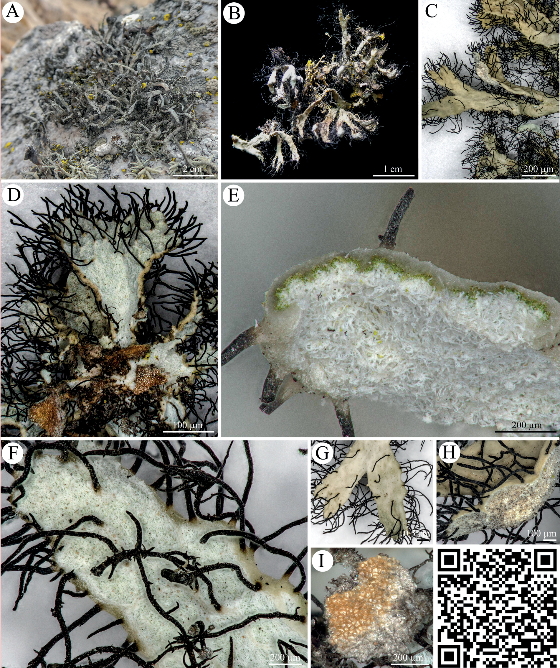
Heterodermia follmannii A, B photographs of Heterodermia follmannii with its gray thallus and black cilia C top view of thallus lobes D bottom view of thallus lobes without lower cortex showing the white, loose medulla and brownish attachment sides E cross section of thallus lobe showing the photobiont layer and the gray upper cortex which forms a rim on the ventral side with an open medulla F ventral side of thallus lobe with open medulla and black, long and branched cilia G top view H upwards bent terminal thallus part which is slightly bloated I ventral view of the attachment side. QR code redirects to 3D scan of Heterodermia follmannii. PW: Lichen. |