|
||
|
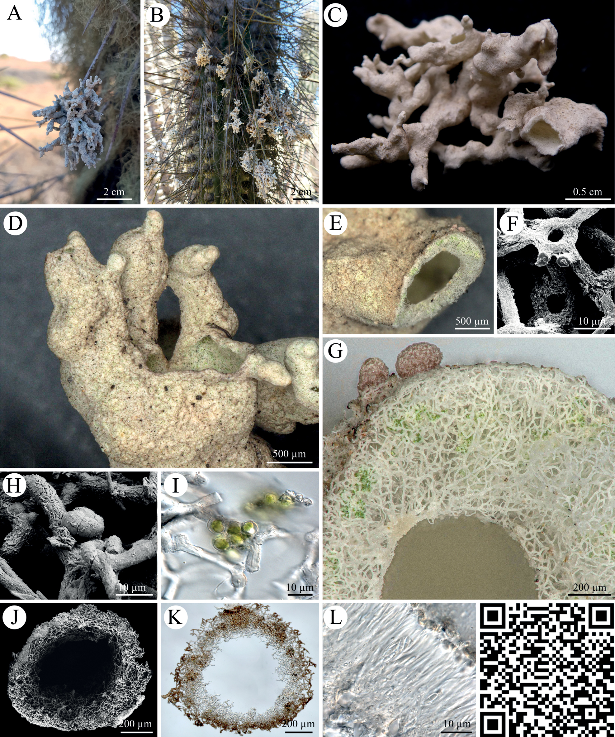
Roccellinastrum spongoideum A, B photographs showing R. spongoideum on the downfacing cactus needles of different Eulychnia cacti in the National Park Pan de Azucar C photograph of byssoid lichen thallus D, E close-up of byssoid thallus showing the coarse hyphal structure F SEM image of hyphal loops G close-up of a cross section with a loose hyphal network, patchy arrangement of photobionts and pink apothecia H SEM image of four ‘micareoid’ photobionts on fungal hyphae I light microscopy of micareoid photobionts with large vacuole-like structures J, K SEM image and light microscopy of the lichen thallus L microscopic cross section through apothecium with two-celled spores divided by a septum. QR code redirects to 3D scan of R. spongoideum. PW: Lichen. |