|
||
|
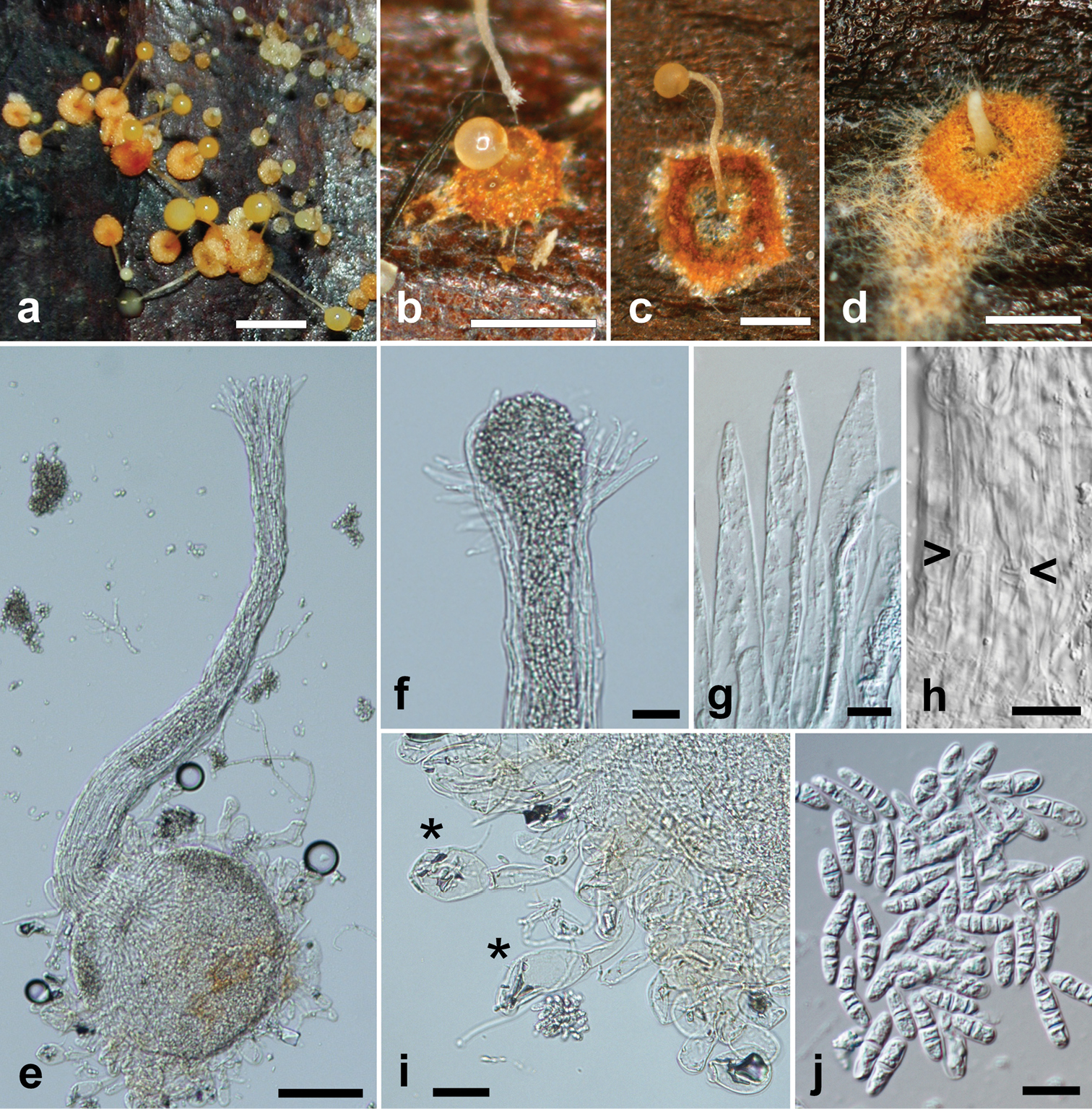
Pycnopulvinus aurantiacus (holotype PUL F2679). a Field photo of fresh sporocarps on palm leaf. Note the variable size and color of the sporocarps. Bar = 2 mm b–d Sporocarps of various stages and sizes after drying. Bars = 0.5 mm e Sporocarp as seen under the light microscope. Note the swollen pigmented base with surrounding large globose cells and the long tubular neck with a widening tip. Bar = 200 μm f Tip of the neck with spore mass exiting the sporocarp. Bar = 25 μm g Ostiolar hyphae at the tip of the neck. Bar = 10 μm h Outer surface of the neck, wide hyphae are visible, septa are marked with arrows. Bar = 10 μm i Globose cells (marked with asterisk) surrounding the base of the sporocarp. Bar = 25 μm j Multicellular spores produced inside the sporocarps. Note the four-celled spores on the left breaking into smaller compartments. Bar = 10 μm. |